Download
1 / 55
710 likes | 1.19k Vues
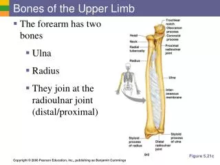
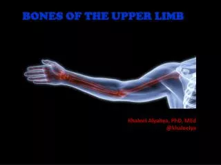
Bones of the Lower Limb. Dr. Fadel Naim Orthopedic Surgeon Faculty of Medicine IUG. The lower limbs specialized for locomotion The primary function of the lower limbs: Support the weight of the body Provide a stable foundation in: Standing Walking Running

E N D
Bones of the Lower Limb Dr. Fadel Naim Orthopedic Surgeon Faculty of Medicine IUG
The lower limbs specialized for locomotion The primary function of the lower limbs: Support the weight of the body Provide a stable foundation in: Standing Walking Running Similar in structure in many respects to the upper limbs Have less freedom of movement The upper limb is united to the trunk by only a small joint, (the sternoclavicular joint) The two hip bones articulate: Posteriorly with the trunk at the strong sacroiliac joints Anteriorly with each other at the symphysis pubis. The lower limbs are more stable
Organization Of The Lower Limb • The lower limbs are divided into different regions and compartments • The regions: • The gluteal region • The thigh • The knee • The leg • The ankle • The foot • The thigh and the leg are compartmentalized • Each compartment with own muscles • Perform group functions • Own distinct nerve and blood supply
The bone of lower limb ilium pubis ischium The pelvic girdle: hip bone The bone of free lower limb femur patella tibia fibula foot tarsal bones metatarsal bones phalanges of foot
Bones Of The Gluteal Region Hip bone • The ilium • The ischium • The pubis • Meet one another at the acetabulum • Articulate with the sacrum at the sacroiliac joints • Form the anterolateral walls of the pelvis • Articulate with one another anteriorly at the symphysis pubis.
Ilium Upper 2/5 Ischium Posterior/lower 2/5 Pubis Anterior/lower 1/5
The Ilium • The upper flattened part of the hip bone • The iliac crest • Can be felt through the skin along its entire length • Anterior superior iliac spine • Anterior inferior iliac spine • Posterior superior iliac spine • posterior inferior iliac spine. • The iliac tubercle lies about 5 cm behind the anterior superior spine. • Greater sciatic notch
Theischium • L shaped • The body • The ramus • The ischial spine • Projects from the posterior border of the ischium and intervenes between the greater and lesser sciatic notches. • The ischial tuberosity
Thepubis • Divided into: • A body • A superior ramus • An inferior ramus • The bodies of the two pubic bones articulate with each other in the midline anteriorly at the symphysis pubis • The superior ramus joins the ilium and ischium at the acetabulum • The inferior ramus joins the ischial ramus below the obturator foramen.
Anterior superior iliac spine Iliac crest Ilium Anterior inferior iliac spine Pubis Acetabulum Ischium Obturator foramen Ischial tuberosity Anterior
TheAcetabulum • On the outer surface of the hip bone is a deep depression, called the acetabulum • Articulates with the head of the femur to form the hip joint • The inferior margin of the acetabulum is deficient and is marked by the acetabularnotch • The articular surface of the acetabulum is limited to a horseshoe shaped area and is covered with hyaline cartilage. • The floor of the acetabulum is non-articular and is called the acetabular fossa
sacroiliac joint Sacrum ilium Pelvic Girdle iliac fossa ischium acetabulum obturator foramen pubis pubic symphysis
iliac crest HIP BONE anterior superior iliac spine greater sciatic notch ischial spine lesser sciatic notch ischial tuberosity
Femur • Articulates above with the acetabulum and below with the tibia and the patella • The upper end of the femur has • A head • A neck • Greater and lesser trochanters • Thehead forms about two thirds of a sphere • Articulates with the acetabulum of the hip bone to form the hip joint
The Neck of the Femur • Connects the head to the shaft • Pass downward, backward, and laterally • Makes an angle about 1250 with the long axis of the shaft. • (Slightly less in the female)
The Greater And Lesser Trochanters • Large eminence situated at the junction of the neck and the shaft • Connecting the two trochanters are the intertrochanteric line anteriorly • Prominent intertrochanteric crest posteriorly
Linea Aspera • The shaft of the femur is smooth and rounded on its anterior surface but posteriorly has a ridge, the linea aspera • Attachment of muscles and intermuscular septa. • The margins of the linea aspera diverge above and below.
The medial margin continues below as the medial supracondylar ridge to the adductor tubercle on the medial condyle • The lateral margin becomes continuous below with the lateral supracondylar ridge. • The shaft becomes broader toward its distal end and forms a flat, triangular area on its posterior surface called the popliteal surface
The lower end of the femur has lateral and medial condyles, separated posteriorly by the intercondylar notch. • The anterior surfaces of the condyles are joined by an articular surface for the patella. • Above the condyles are the medial and lateral epicondyles • The adductor tubercle is continuous with the medial epicondyle.
Fovea of femoral head Femoral head The femur Greater trochanter Neck of femur Lesser trochanter Intertrochanteric line Shaft of femur Lateral epicondyle Medial epicondyle Patellar surface Anterior view
Intertrochanteric crest Gluteal tuberosity Linea aspera Intercondylar fossa Medial condyle Lateral condyle Posterior view
PATELLA • The largest sesamoid bone • Triangular • Its apex lies inferiorly • The posterior surface articulates with the condyles of the femur
Tibia • The large weight-bearing medial bone of the leg • It articulates with: • The condyles of the femur • The head of the fibula • The talus • The distal end of the fibula • It has an expanded upper end, a smaller lower end, and a shaft.
Tibia • At the upper end: • The lateral and medial condyles (sometimes called lateral and medial tibial plateaus), • Articulate with the lateral and medial condyles of the femur • Anterior and posterior intercondylar areas separate the upper articular surfaces of the tibial condyles • Intercondylar eminence lies between these areas
The lateral condyle possesses on its lateral aspect a small circular articular facet for the head of the fibula. • The medial condyle has on its posterior aspect the insertion of the semimembranosus muscle
Medial tibial condyle Lateral tibial condyle Fibular head Tibial tuberosity
The Shaft Of The Tibia • Triangular in cross section • Three borders and three surfaces • Anterior, medial borders and the medial surface are subcutaneous. • The anterior border is prominent and forms the the shin. • At the junction of the anterior border with the upper end of the tibia is the tuberosity, • Receives attachment of the ligamentum patellae. • The anterior border becomes rounded below, where it becomes continuous with the medial malleolus
The Shaft Of The Tibia • The lateral or interosseous border gives attachment to the interosseous membrane • The posterior surface of the shaft shows an oblique line, the soleal line for the attachment of the soleus muscle
The lower end of the tibia is slightly expanded and on its inferior aspect shows a saddle-shaped articular surface for the talus. • The lower end is prolonged downward medially to form the medial malleolus. • The lateral surface of the medial malleolus articulates with the talus. • The lower end of the tibia shows a wide, rough depression on its lateral surface for articulation with the fibula.
Tibia medial condyle medial malleolus anterior crest tibialtuberosity lateral condyle
FIBULA • The slender lateral bone of the leg • No part in the articulation at the knee joint • Below it forms the lateral malleolus of the ankle joint. • No part in the transmission of body weight • Provides attachment for muscles. • An expanded upper end, a shaft, and a lower end.
The upper end, or head • A styloid process. • Articular surface for articulation with the lateral condyle of the tibia • The shaft of the fibula • Long and slender. • Four borders and four surfaces • The medial or interosseous border gives attachment to the interosseous membrane.
The lower end of the fibula • Forms the triangular lateral malleolus, which is subcutaneous. • On the medial surface of the lateral malleolus is a triangular articular facet for articulation with the lateral aspect of the talus.
Fibula lateral malleolus head Patella
Bones of the Foot • The bones of the foot are: • The tarsal bones • The metatarsals • The phalanges
Right foot phalanges Superior view metatarsals tarsals steven lee M.S. Pathology FTCC
TARSAL BONES • The tarsal bones are • The calcaneum • The talus • The navicular • The cuboid • The three cuneiform bones. • Only the talus articulates with the tibia and the fibula at the ankle joint
Calcaneum • The largest bone of the foot • The prominence of the heel • It articulates • above with the talus • in front with the cuboid.
Talus • The talus articulates: • Above at the ankle joint with the tibia and fibula • Below with the calcaneum • In front with the navicular bone. • It possesses • A head • A neck • A body • Numerous important ligaments are attached to the talus • No muscles are attached to this bone.
Navicular Bonescaphoid bone • Is located on medial side of the foot between talus and the three cuneiforms. • It articulates posteriorly with talus and anteriorly with the 3 cuneiforms.
Cuboid Bone • The cuboid bone is placed on the lateral side of the foot, in front of the calcaneus, and behind the fourth and fifth metatarsal bones.
Cuneiform Bones • The three small, wedge-shaped • Articulate proximally with the navicular bone and distally with the first three metatarsal bones. • Their wedge shape contributes greatly to the formation and maintenance of the transverse arch of the foot
METATARSAL BONES AND PHALANGES • Resemble the metacarpals and phalanges of the hand • Each possesses a head distally, a shaft, and a base proximally • The five metatarsals are numbered from the medial to the lateral side. • The first metatarsal bone is large and strong and plays an important role in supporting the weight of the body. • The head is grooved on its inferior aspect by the medial and lateral sesamoid bones in the tendons of the flexor hallucis brevis.
METATARSAL BONES AND PHALANGES • The fifth metatarsal has a prominent tubercle on its base that can be easily palpated along the lateral border of the foot. • The tubercle gives attachment to the peroneus brevis tendon. • Each toe has three phalanges except the big toe, which possesses only two.