BONES OF THE UPPER LIMB
400 likes | 845 Vues
BONES OF THE UPPER LIMB . Dr. Khaleel Alyahya Assistant Professor College of Medicine King Saud University. Dr. Jameela Al- Medany Associate Professor College of Medicine King Saud University. OBJECTIVES. At the end of the lecture, students should:

BONES OF THE UPPER LIMB
E N D
Presentation Transcript
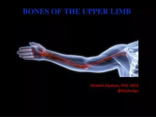
BONES OF THE UPPER LIMB Dr. Khaleel Alyahya Assistant Professor College of Medicine King Saud University Dr. Jameela Al-Medany Associate Professor College of Medicine King Saud University
OBJECTIVES At the end of the lecture, students should: • List the different bones of the Upper Limb. • List the characteristic features of each bone. • Differentiate between bones of right and left sides. • List the articulations between the different bones.
Bones of Upper Limb It consists of the following: Pectoral Girdle Arm Forearm Hand
Pectoral Girdle • It composed of Two bones: • Clavicle • Scapula • It is very light and it allows the upper limb to have exceptionally free movement.
Clavicle • It is a long bone lying horizontally across the root of the neck • It is subcutaneous throughout its length. • Functions: • Holds the arm away from the trunk. • Transmits forces from the upper limb to the axial skeleton. • Provides attachment for muscles. • If the clavicle is broken, the whole shoulder region caves in medially.
Clavicle • Its medial 2/3 is convex forward • Its lateral 1/3 is concave forward • Its medial (sternal) end is rounded. • Its lateral (acromial) end is flattened. • It has two surfaces: • Superior • Inferior • The Inferior surface shows: • Conoid tubercle & Trapezoid line.
Articulations • Medially, sternoclavicular joint • with the Manubrium • Laterally, Acromioclavicular joint • with the Acromial end of the scapula • Inferiorly, costoclavicular Joint • with the 1st rib
Scapula • It is a triangular flat bone. • Extends between the 2nd _ 7th ribs. • It has: • Two surfaces: Anterior & Posterior. • Three Borders: superior, medial (vertebral) & lateral (axillary). • Three Processes: Spine, Coracoid & Acromion. • Three Angles: superior, lateral (forms the Glenoid cavity) & inferior. • Suprascapular notch: medial to coracoid process. • It is a nerve passageway.
Fossae of Scapula • It has Three Fossae: • Two on the posterior surface: • Supraspinous Fossa • above the spine. • Infraspinous Fossa • below the spine. • One on the Anterior surface: • Subscapular Fossa.
Arm (Humerus) • It is formed by a single bone. • It is a typical long bone. • It has: • Upper End: • Head: Smooth& forms 1/3 of a sphere. • Anatomical neck: Immediately below the head. • Greater & Lesser tubercles: separated by Intertubercular Groove. • Surgical Neck: between the upper end and the shaft. surgical
Arm (Humerus) • Shaft: • Anterior & Posterior surfaces. • Deltoid tuberosity: • A rough elevation halfway down the lateral aspect. • Spiral (Radial) groove: • Runs obliquely down the posterior aspect of the shaft. • It lodges the important radial nerve & vessels. surgical
Arm (Humerus) • Distal End: • Medial : Trochlea. • Lateral: Capitulum. • Above the trochlea (on the anterior surface): Coronoid fossa. • Above the trochlea (on the posterior surface): Olecranon fossa. • Above capitulum: • Radial fossa. • Epicondyles: medial & lateral. surgical
Articulations • Head of the humerus with the glenoid cavity of the scapula form the Shoulder joint. • Lower end (Trochlea & Capitulum) with the upper ends of the radius & ulna form theElbow joint.
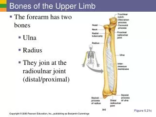
Forearm • Formed of two bones: • The Radius is the lateral bone. • The Ulna is the medial bone.
Radius • Proximal (Upper)) end: • Head: small & circular& Its upper surface is concave. • Neck • Radial (Biciptal) Tuberosity • Shaft: • Wider below than above • Three surfaces: Anterior, Lateral & Posterior • Sharp medial interosseous border. • Pronator tubercle: halfway down the lateral side. • Distal (Lower) end: • Styloid process: laterally. • Ulnar notch: medially. • Dorsal tubercle: posteriorly.
Ulna • Proximal (Upper)) end: • Posterior: Olecranon process • Forms the prominence of the elbow. • Anterior: Coronoid process. • Both they are separated by the Trochlear notch • Lateral : Radial notch • Shaft: • Three surfaces (Anterior, Medial & Posterior). • Sharp lateral interosseous border. • Distal (Lower) end: • Small rounded head • Medial : Styloid process • Lateral : Radial notch
Articulations of Radius & Ulna • Distal end of Humerus with the proximal ends of Radius & Ulna Elbow joint • Proximal Radioulnarjoint • Distal Radioulnarjoint • The two bones are connected by the flexible interosseous membrane Proximal Radioulnar joint.
Hands • The skeleton of the hand consists of the: • Carpalsfor the carpus (wrist) • Metacarpalsfor the palm • Phalangesfor the fingers
Carpal Bones • Eight carpal bones arranged in two irregular rows, each of four. • They present Concavity on their Anterior surface. • Proximal row (from lateral to medial): • Scaphoid • Lunate • Triquetrum • Pisiform • Distal row (from lateral to medial): • Trapezium • Trapezoid • Capitate • Hamate
Metacarpals& Phalanges Bones • Metacarpals: • Five bones, each has a Base, Shaft, and a Head. • They are numbered 1-5 from the thumb. • The 1st metacarpal is the shortest and most mobile. • When the fist is clenched, the heads of the metacarpals become obvious (knuckles) • Phalanges : • Each hand contains 14 phalanges. • Three in each finger (proximal, middle & distal) except in the thumb which has only two (proximal &distal).
Articulations • Bases of the Metacarpal bones articulate with the distal row of the carpal bones • Carpometacarpal joints • Heads (knuckles) articulate with the Proximal Phalanges • Metacarpophalangeal joints • The phalanges articulate with each other • Interphalangeal joints • Distal end of Radius with the Proximal Raw of Carpal bones • Wrist joint