1 / 0

Traumatic Brain Injury Re-Visited: A Pictorial Review

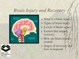
Traumatic Brain Injury Re-Visited: A Pictorial Review. Abstract #: eEdE-70. Cedric Pluguez-Turull, MD Wilmarie Rivera, MD Stephanie Baussan , MD Jose Lara, MD Cristina Quintero-Estades, MSIV Jose A. Quintero- Estades , MSIII Sean Maldonado, MSIII Eduardo Labat , MD.

zora
Télécharger la présentation

Traumatic Brain Injury Re-Visited: A Pictorial Review

An Image/Link below is provided (as is) to download presentation Download Policy: Content on the Website is provided to you AS IS for your information and personal use and may not be sold / licensed / shared on other websites without getting consent from its author. Content is provided to you AS IS for your information and personal use only. Download presentation by click this link. While downloading, if for some reason you are not able to download a presentation, the publisher may have deleted the file from their server. During download, if you can't get a presentation, the file might be deleted by the publisher.

E N D

Presentation Transcript


  1. Traumatic Brain Injury Re-Visited: A Pictorial Review

    Abstract #: eEdE-70 Cedric Pluguez-Turull, MD Wilmarie Rivera, MD Stephanie Baussan, MD Jose Lara, MD Cristina Quintero-Estades, MSIV Jose A. Quintero-Estades, MSIII Sean Maldonado, MSIII Eduardo Labat, MD
  2. Disclosure of Commercial Interest Neither us nor any of our immediate family members have a financial relationship with a commercial organization that may have a direct or indirect interest in the content.
  3. Objectives Introduce the concept of of traumatic brain injury. Provide a review of the indications for radiological evaluation and the most common associated complications of brain trauma. Review the pathophysiology, mechanism of injury (MOI), characteristic imaging findings, management strategies and classic signs of traumatic brain injuries by examining computed tomography and MR imaging findings.
  4. Introduction Traumatic brain injuries (TBIs) are a very common cause of hospital admission following trauma, and are associated with significant long-term morbidity and mortality. Every year, at least 1.7 million TBIs occur either as an isolated injury or along with other injuries. TBI contributes as much as a third (30.5%) of all injury-related deaths in the United States.
  5. White Matter Skull Normal Brain Anatomy Subarachnoid Space Prefrontal Area Corpus Callosum Lateral Ventricles Lateral Ventricles Basal Ganglia Caudate Nucleus Putamen Temporal Lobe Globus Pallidus Third Ventricle Grey Matter Sulcus Occipital Lobe Pons Gyrus Falx Sub-Arachnoid Space Inferior horn of Lateral Ventricle Interpeduncular Cistern
  6. Diagnostic Imaging Appropriateness Subacute / Chronic Head Injury Acute Head Injury Penetrating Closed Closed No neuro deficit Minor or mild trauma (GCS>13), no risk factors, no neuro deficit Rule out carotid or vertebral artery dissection Minor or mild trauma (GCS>13), focal neuro deficit and/or risk factors OR Moderate to Severe trauma Cognitive and/or Neuro deficit CT Head w/o contrast OR CTA Head and Neck with contrast CTA Head and Neck with contrast OR MRA Head and Neck without contrast*** OR MRA Head and Neck wwo contrast***, MRI Head w/o contrast (Add DWI), OR CT Head w/o contrast CT Head w/o contrast* MRI Head w/o contrast CT Head w/o contrast OR MRI Head w/o contrast** Skull Fracture CT Head w/o contrast [CTA Head and Neck with contrast may be appropriate] * Usually appropriate, but is low yield. ** May be appropriate, but only used for problem solving. DWI helpful, especially non-accidental trauma in < 2y/o . *** Add T1 Neck view to study
  7. Overview Intracranial Bleeding Microhemorrhages Epidural Hematoma Subdural Hematoma Subarachnoid Hemorrhage (SAH) Intraventricular Hemorrhages Skull Fractures Linear Depressed Basilar Compound Parenchymal Injury Parenchymal contusion Diffuse Axonal Injury (DAI) Penetrating Injury Laceration Foreign Body Secondary Effects Edema Hypoxic-ischemic encephalopathy Cerebral herniation Post-traumatic Encephalocoele Pneumocephalus Post-Traumatic Encephalomalacia
  8. Intracranial Bleeding: Hemorrhages and Hematomas Subdural Hematoma Epidural Hematoma Microhemorrhages Intraparenchymal Hematoma Intraventricular Hemorrhage Contusion
  9. Traumatic Microhemorhages Epidemiology: In patients with hemorrhagic conditions (to include trauma) or ischemic cerebrovascular disease (up to 60%). Microhemorrhages often seen in hypertensive patients (up to 56%), DAI and can be seen in healthy, asymptomatic populations (up to 6.4%). Mechanism: Caused by rupture of small blood vessels in subcortical white matter or basal ganglia. Associated with hemosiderin deposition from earlier hemorrhagic episodes. Number and location of lesions can be associated with cognitive dysfunction. May anticipate the occurrence of intracranial hemorrhage or lacunar infarction. Management: Typically asymptomatic and require no intervention. Complications: Depends on underlying pathology. Imaging Findings: MRI > CT for detection. NECT: punctate hyperdensities MRI: Ovoid-shaped foci of decreased signal intensity measuring < 5mm. Best seen on T2 weighted sequences, such as gradient-echo MRI, and often not seen with other modalities. Characteristically seen as punctate regions with signal loss (darkening). Classic signs: Punctate regions with signal loss on T2WI MRI.
  10. Traumatic Microhemorhages (a) (b) Fig 1. Nonenhanced Head CT of different trauma patients (a), (b) show small foci of hemorrhage involving the juxtacortical white matter (yellow arrowhead). Ventriculostomy catheter is also noted in the left frontal parafalcineparenchyma (red arrow) in patient (b).
  11. Epidural Hematoma Epidemiology: Trauma most common. Found in 1-4% of imaged head trauma patients, 5-15% of patients with fatal head injuries. More common in patients < 20 years and in males (M:F = 4:1). Uncommon in elderly and infants. More than 95% are unilateral. Supratentorial (90-95%), temporo-parietal (65%). Mechanism: Blunt trauma to head. Arterial (90%) > Venous (10%). Arterial EDH most often near middle meningeal artery groove fracture; venous EDH related to fractures near dural sinus attachments. Management: Nearly always requires surgical evacuation. EDH < 1 cm thick may be managed non-operatively if no concomitant cerebral edema. May consider endovascular adjunct treatment and repeat CT in first 36 hours to monitor for change. Complications: Mass effect causing herniation, skull fracture in 95% of patients, cerebral contusions and countercoup subdural hematoma.
  12. Epidural Hematoma Imaging Findings: Best imaging tool is NECT. CT: NECT- Acute: 2/3 hyperdense, 1/3 mixed density. Low-density "swirl" sign means active/rapid bleeding with unretracted clot. Medial hyperdense margin represents displaced dura. Air in EDH (20%) suggests sinus or mastoid fracture. Bilateral: "hourglass" brain. Chronic: hypo/mixed density. CECT- Chronic: Peripheral dural enhancement from neovascularization and granulation. Classic signs: Hyperdense biconvex extra-axial collection on NECT. CT "comma" sign - EDH plus subdural hematoma MRI: T1WI - Acute: isointense. Subacute/early chronic: hyperintense. Black line between EDH and brain: displaced dura. T2WI - Acute: variable hyper to hypointense. Early subacute: hypointense. Late subacute/early chronic: hyperintense. Black line between EDH and brain: displaced dura. T1WI C+ - Venous EDH: Displaced dural sinus by hematoma. MRV: Used to assess for patency of venous sinus. Will show displaced venous sinus flow by hematoma.
  13. Epidural Hematoma Fig 2B Fig 2A Fig 2A. Non-enhanced Head CT shows right frontal lenticular extra-axial hyperdense fluid collection (yellow arrowhead) consistent with epidural hematoma. Fig 2B. Non-enhanced Head CT shows right middle cranial fossa lenticular extra-axial hyperdense fluid collection (yellow arrowhead) consistent with epidural hematoma. Note associated displaced right sphenoid bone fracture (red arrow) and countercoup left anterior temporal contusion.
  14. Subdural Hematoma (SDH) Epidemiology: Caused by head trauma, typically from MVA and falls. 11% of mild to moderate head injuries and in 20% of severe head injuries. Predisposing factors include: cerebral atrophy, chronic alcoholism, previous TBI, advanced age and use of anti-thrombotic agents. M >F and average age 31 to 47. Mortality for patients with SDH requiring surgery can be as high as 60%. Mechanism: Results from closed-head injury that tear bridging veins that drain from the brain’s surface to the dural sinuses. Management: Small SDH (clot thickness < 10mm and midline shift <5mm) in stable patients may be monitored and managed conservatively, otherwise craniotomy. Lumbar puncture and anticoagulation is contrandicated. Complications: Cerebral herniation, elevated intracranial pressure and chronic SDH: hygroma (over 50% will grow in size).
  15. Subdural Hematoma (SDH) Imaging Findings: Best imaging tool is NECT NECT: Hyperdense crescent shaped lesion along the hemisphere. Subacute and chronic SDH appear iso- or hypodense; collagen envelope may enhance with contrast. May present with mass effect. MRI: Greater sensitivity than CT; used to follow up unclear or negative CT’s. Acute clots are hypointense on T2 images. Over the course of weeks, the signal becomes bright on T1 and T2 images due to the presence of methemoglobin. After several months, the clot becomes hypointense on T1 owing to the hemosiderin remnant. Classic Signs: Hyperdense “crescent shaped” extra-axial lesion that crosses sutural margins on NECT. Fig 3. Non-enhanced Head CT shows left panhemispheric hyperdense crescent shaped extra-axial fluid collection (yellow arrow) consistent with acute subdural hematoma. Note effacement of adjacent sulci due to mass effect.
  16. Subdural Hematoma (SDH) Fig 4A Fig 4B Fig 4A. Axial images of non-enhanced Head CT shows right panhemispheric crescent shaped extra-axial fluid collection (yellow arrowhead) of intermediate attenuation consistent with subacute subdural hematoma. Fig 4B. Coronal images of non-enhanced Head CT show hyperdense right parafalcine and supratentorial subdural hemorrhage (yellow arrowhead).
  17. Traumatic Subarachnoid Hemorrhage (tSAH) Epidemiology: In 40% to 61% of cases of TBI. Mortality can be as high as 50%. Bleeding patterns and location can distinguish traumatic SAH from more common causes, such as aneurysm rupture. Mechanism: Hemorrhage may result from laceration of cortical vessels, spread from intraventricular hemorrhage, or direct extension from cortical contusions. Most trauma patients exhibit blood in the superficial cortical sulci. Blood is released into the CSF with arterial pressure, which increases ICP and compromises brain oxygen delivery. Management: Intubation if GCS < 8 or hemodynamically unstable. Thrombosis prophylaxis, IVF and calcium channel blockers. Complications: Rebleeding most commonly within the first 24hrs., increased ICP (>50%), cerebral ischemia (50% - most commonly caused by vasospasm), seizures (18%) and hydrocephalus (15%).
  18. Traumatic SAH Imaging Findings: Best imaging tool is NECT. NECT with thin cuts is the preferred method for diagnosis, with a sensitivity of almost 100% within the first 6-12 hours following TBI. High-attenuation fluid in SA space. Most evident in largest SA spaces such as suprasellar cistern and sylvian fissures. Clots can be seen in subarachnoid space before 24 hours. Intracerebral extension in over 20% of patients and intraventricular blood in over 15% of patients. Classic Signs: NECT – high-attenuating SA spaces after acute head trauma. Fig 5. Non-enhanced Head CT shows hyperdense blood within of the bilateral frontal upper convexity subarachnoid spaces (yellow arrowheads); consistent with acute subarachnoid hemorrhage.
  19. Traumatic SAH (d) (c) (a) (b) Fig 6. Axial non-enhanced CT (a) and T1W images (b) show evident left parafalcine/tentorial subdural hemorrhage (yellow arrowheads) and left anterior temporal hemorrhagic contusion (red arrow). No definite subarachnoid hemorrhage is noted. Axial FLAIR (c) and (d) images show lack of suppression of subarachnoid CSF in the occipital lobes (yellow arrowheads); consistent with post-traumatic subarachnoid hemorrhage.
  20. Intraventricular Hemorrhage (IVH) Epidemiology: Present in 60% of patients with corpus callosal DAI, 12% in patients without. Most common in 15–24 year olds. More common in men. Mechanism: Most commonly results from disruption of subependymal veins. Other causes include bleeding from choroid plexus, shearing injuries and basal ganglia/intracerebral hemorrhage with rupture into ventricles. Isolated IVH in absence of parenchymal hematoma is unusual. Management: Ventriculostomy has excellent results following r-TPA thrombolytic therapy. Repeat NECT to evaluate for complications. Complications: DAI, SAH, cerebral contusion, hydrocephalus and seizures.
  21. Intraventricular Hemorrhage (IVH) Imaging Findings: Best imaging tool - NECT > MR CT: NECT - Hyperdense intraventricular blood that may fill and even expand ventricle. Fluid-heme level common. MRI: T1WI - Fluid-heme level common. T2WI - Fluid-heme. FLAIR - Detection of hemorrhage comparable to CT in acute stage. Classic signs: Hyperdense intraventricular CSF on NECT, fluid-heme level common. Fig 7. Non-enhanced Head CT of two trauma patients shows hyperdense blood within of the anterior aspect of the right temporal horn (a) and layering in the right occipital horn (b) (yellow arrowheads); consistent with acute intraventricular hemorrhage.
  22. Skull Fractures Linear Fracture Depressed Fracture Comminuted Fracture
  23. Skull Fractures Epidemiology: Skull fractures are present in 80% of fatal injuries at autopsy. Simple linear fracture is most common. Basilar skull fractures are 19-21% of all skull fractures. Depressed fractures are more often frontoparietal (75%), but can also be temporal (10%), occipital (5%), or other (10%). Depressed fractures are compound fractures in 75-90% of cases. Mechanism: In infants: neglect, fall, or abuse. In children: falls and bicycle accidents. In adults: motor vehicle accidents or violence. Linear fractures are associated to low-energy blunt trauma over wide surface area of skull. Depressed fractures are associated to high-energy direct blow to small surface area with blunt object.
  24. Skull Fractures Management: Most skull fractures, even depressed, do not require surgery. Surgery in: hematoma, >8-10mm depression, persistent CSF leak, ossicle disarticulation or increased ICP. Antibiotics for contaminated fractures Complications: Epidural or subdural hematoma, lacerated dura or arachnoid, parenchymal injury, CSF leak, delayed CN deficits, infarct, hearing loss, persistent vertigo, and post-traumatic encephalocele. Traumatic carotid cavernous fistula is a serious complication associated to basilar fractures (3.8%) that if untreated can lead to blindness. Fig 8. Non-enhanced bone window axial Head CT images unzoomed (a) and zoomed in (b) show linear non-depressed fracture of the right parietal bone (yellow arrow).
  25. Skull Fractures Imaging Findings: Best imaging tool is NECT; thin-slice high-resolution if skull base involved. Add MR if depressed fracture. Plain films have no role Perform CTA or DSA if traumatic carotid cavernous fistula is suspected. Bone CT: Linear fracture: Sharply delineated lucent line. Depressed fracture: Fragment displaced inward. Compound: Air in intracranial region that may be associated to soft tissue swelling. Basilar fracture: Pneumocephalus common, air-fluid level within adjacent air cell, nasal cavity fluid if CSF rhinorrhea is present, ear cavity fluid from CSF otorrhea or blood density from hemotympanum and air in TMJ glenoid fossa may be only CT sign of inconspicuous skull base fracture. CTA:quickly evaluate for vascular injury if carotid canal involved. Better than MRA. MRI: T2WI - Best to delineate dural injury, FLAIR: Hyperintense cerebral contusion, & T2* GRE: Foci of hemorrhage susceptibility. Classic Signs: Linear calvarial lucency on radiography.
  26. Parenchymal Injury Diffuse Axonal Injury (DAI) with Microhemorrhages Parenchymal Laceration Contusion Foreign Body in Penetrating Trauma
  27. Parenchymal Contusion Epidemiology: Annual incidence is 200 per 100,000 brain trauma-related hospitalizations. Contusion is the 2nd most common primary traumatic neuronal injury (44%); DAI is most common. Mechanism: Results from stationary head struck by object, which results in direct injury beneath impact site. Contusion is rare without fracture. Management: Central goal is to prevent and treat secondary injury. Mass effect and herniation may require surgical evacuation. Complications: Herniations and/or mass effect with secondary infarction. Hydrocephalus due to hemorrhage. Others include hypoxia, hypotension, ischemia, brain edema, and increased intracranial pressure.
  28. Parenchymal Contusion Imaging Findings: Best imaging tool is CT to detect acute hemorrhagic contusions, other intracranial lesions, and herniations. MR to detect presence and delineate extent of lesions: FLAIR for edema and SAH, and GRE for hemorrhagic foci. NECT: Early: Patchy, ill-defined, low-density edema with small foci of hyperdense hemorrhage. 24-48 hours - Edema, hemorrhage, and mass effect often increase. New foci of edema and hemorrhage may appear and petechial hemorrhage may coalesce. Sub-Acute/Chronic - Become isodense, then hypodense. Eventual encephalomalacia with volume loss. Perfusion CT: more sensitive than NECT in detection of cerebral contusions (87.5% vs. 39.6%). MRI: T1WI - Acute: Inhomogeneous isointensity and mass effect. Chronic: Focal or diffuse atrophy. FLAIR - Acute: Best for hyperintense cortical edema and subarachnoid hemorrhage (SAH). Chronic - Hyperintense demyelination and microglial scarring. Hypointense hemosiderin staining. T2* GRE - Acute: Hypointense hemorrhagic foci "bloom”. Chronic: Hypointense hemosiderin deposits. DWI - Hyperintense in areas of cell death with ADC decreased. Isointense in vasogenic edema with increased ADC. DTI: white matter damage in minor head trauma when CT and routine MR are normal. Classic signs: Patchy hemorrhages within edematous background.
  29. Parenchymal Contusion Fig 9A Fig 9B Fig 9A. Non-enhanced axial Head CT images show bifrontal basal multifocal acute hemorrhagic contusions (yellow arrowheads); which are common due to rough surface of the anterior cranial fossa. Fig 9B. Non-enhanced axial Head CT images shows right temporal acute hemorrhagic contusion (yellow arrowheads) with minimal surrounding parenchymal edema and slight mass effect.
  30. Diffuse Axonal Injury (DAI) Epidemiology: Most common in young men. Often have coexisting injury following trauma (such as a contusion). Mechanism: Most frequently caused by shearing mechanisms from TBI. Common mechanisms include rapid acceleration/deceleration, direct impact, and blast waves. Mechanical forces result in shearing of white matter tracts as well as cerebral edema. Management: Minimize secondary damage related to cerebral hypoxia or edema. Complications: Related to the mechanism of injury and location of TBI (e.g. subdural hematoma). Long-term include: focal neurologic, cognitive deficits or vegetative state. Imaging Findings: Multiple small lesions seen along the gray-white matter junction. NECT: Lesions are rarely visible on CT unless they are also hemorrhagic. Hemorrhagic lesions will have hyperdense foci, microhemorrhages most common. Nonhemorrhagic DAI will appear hypodense and is detectable in under 20% of cases. MRI: MRI is the modality of choice, capable of detecting up to 92% of DAI lesions with T2 weighted imaging, even in the absence of hemorrhage. White matter lesions will appear bright on T2 imaging with surrounding edema. Classic Sign: Multiple small T2 hyperintense lesions along gray-white matter junction on MRI.
  31. Diffuse Axonal Injury (DAI) Fig 10. (a), (b) Non-enhanced Head CT of trauma patient shows multiple small hemorrhagic foci (yellow arrowheads)in the right frontal lobe subcortical white matter, right genu and splenium of the corpus callosum (a) more evident on follow up exam(b); with interval development of new left frontal hemorrhagic focus; most consistent with shearing injury. Fig 10 Fig 11. (a), (b) MRI of young trauma patient with persistent obtunded state shows subtle left frontal juxtacortical white matter hyperintensity on FLAIR sequences (a) with corresponding susceptibility artifact on T2* sequences (yellow arrowhead); most consistent with shearing injury given clinical scenario. Fig 11
  32. Foreign Body associated to Penetrating Injury Imaging Findings: Best imaging tool is a NECT. CT: NECT - Best assessment of extent of soft tissue injury. Identify entrance and exit wounds. Osseous entry and exit sites and pneumocephalus shown to better advantage. Metallic fragments are easier to evaluate. MRI: T1WI and T2W1: Variable signal including hemorrhage, foreign bodies or air. T2WI may show edema from pressure wave. T2* GRE: "Blooming" from hemorrhage, as well as susceptibility artifact from foreign bodies, air. DWI: Secondary infarction. CTA and MRA: Evaluate for pseudoaneurysm, dissection, or traumatic AV fistula. MRV: Evaluates venous injury or thrombosis if missile tract crosses or tears interdural veins or lacerates sinus. Classic signs: Single or multiple intracranial foreign bodies, missile tract, pneumocephalus, and entry wound with or without exit wound. Epidemiology: As in penetrating head injury. Mechanism: Results from cranial trauma from high-velocity projectile, typically gunshot wound. Also from impalement with sharp object, such as stabbing wounds. Management: Dependent upon type and degree of injury but most often includes: Debridement - penetrating objects may be left in place, decompressive craniectomy, CSF diversion to control hydrocephalus (especially in setting of infratentorial injury) & intracranial pressure control. Complications: Highly variable depending on type and degree of traumatic injury but includes motor deficits, cranial nerve palsies, visual field defects and post-traumatic seizures.
  33. Foreign Body associated to Penetrating Injury Fig 12. AP Scout (a), B Lateral Scout (b) , and Axial Nonenhanced CT images (c) show a metallic intracranial foreign body in the left occipital lobe from a prior gunshot (black/yellow arrows). Abundant metallic streak artifact is noted.
  34. Subfalcine Herniation TBI Complications and Delayed Effects Post-Traumatic Encephalomalacia Pneumatocoele Uncal Herniation Post-Traumatic Encephalocoele Subfalcine Herniation Transcalvarial Herniation Tonsilar Herniation
  35. Brain Edema Epidemiology: Vasogenic and cytotoxic brain edema are a common consequence of certain TBIs and major cause of ICP. Mechanism: The cranial vault has a fixed volume, thus edematous changes in brain size can compromise the space needed for vascular flow and CSF cushioning. Imaging Findings: Edema will appear hypodense on CT and T1 images, but bright on T2 or FLAIR images. May also note compressed ventricles or effaced sulci due to increased brain matter Brain edema may cause loss of gray / white matter differentiation in CT scans. Management: Conservative, may require surgical decompression Complications: Brain herniation, vegetative state Fig 13. (A) Axial image of NECT shows diffuse loss of cortical sulci with partial effacement of the grey-white matter differentiation; consistent with cerebral edema. (B) Same study at level of basal cisterns shows effacement and increased attenuation of the basal cisterns and Sylvian fissure (yellow arrowheads) with associated loss of grey-white matter differentiation in this patient with diffuse cerebral edema.
  36. Hypoxic-Ischemic Encephalopathy Refers to inflammatory brain injury following TBI caused by hypoperfusion that can be radiologically evaluated. Epidemiology: Limited literature. Imaging Findings: Abnormal signal intensities can be appreciated on T2 scans. CT: cerebral edema, atrophy, white matter lesions, and changes in ventricular size. CT serves as an important tool for ruling out hemorrhagic lesions MRI: hyperintense deep gray matter lesions of basal ganglia and thalami bilaterally are common findings in term infants with hypoxic encephalopathy, while periventricular involvement is more common in pre-term infants Management: Therapeutic hypothermia, oxygen Complications: Neurologic deficit, brain death
  37. Hypoxic-Ischemic Encephalopathy (b) (c) (d) (a) Fig 14. Axial T2 (a), FLAIR (b), T2* (c) and T1 (d) weighted images show increased signal intensity on T2/FLAIR sequences (yellow arrows) in the thalami bilaterally without corrresponding signal abnormality on T1 weighted or heavily T2 weighted images. Fig 15. (a), (c) DWI and ADC (b), (d) images show areas of restricted diffusion involving watershed areas of the brain including the juxtacortical deep white matter, splenium of the corpus callosum and bilateral caudate nuclei/thalami (yellow arrowheads). (a) (b) (c) (d)
  38. Transcalvarial Herniation Subfalcine Herniation Cerebral Herniation Epidemiology: Subfalcine is most common. Unilateral descending transtentorial is 2nd most common. Mechanism: Trauma most common clinical setting. May occur as a direct cause of TB or secondary to hemorrhage or inflammation caused by brain trauma. Management: Mitigate secondary effects. Decompressive craniectomy. Complications: Focal neurologic deficit, altered mental status, midbrain Duret hemorrhage, ischemia or even brain death (if progressive ICP or mass effect). Uncal Herniation Tonsilar Herniation
  39. Cerebral Herniation Imaging: Best imaging tool is NECT for best rapid screen. Add multiplanar MR with DWI and T2* GRE for ischemic and hemorrhagic complications. NECT: Ventricles displaced; sulci and cisterns obliterated MRI: T1WI provides best anatomic definition while T2WI is the best for complications such as edema, infarcts, or hydrocephalus. T2* GRE: Best for hemorrhagic foci, such as Duret hemorrhages. DWI: Secondary ischemia/infarcts. Fig 16. Coronal NECT after recent trauma and known right panhemispheric subacute subdural hematoma with right to left subfalcine herniation (yellow arrow).
  40. Cerebral Herniation Fig 17. (a), (b) Axial NECT (a) of patient with massive posttraumatic right temporal hemorrhagic contusions. There is partial effacement of the left suprasellar cistern (yellow arrowhead). Coronal reconstructions (b) shows left downward tentorial herniation (yellow arrowhead). Fig 18. (a), (b) Axial NECT (a) shows crowding of the foramen magnum (black arrowheads). Sagittal images of same study (b) show crowding of posterior fossa structures with fullness of foramen magnum (yellow arrowhead). Findings are consistent with tonsillar herniation.
  41. Post-Traumatic Encephalocoele Imaging Findings: Best imaging tool is multiplanar MR or thin-section Bone CT with multiplanar reformats. CT: NECT - contrast resolution limits ability to distinguish encephalocele from paranasal sinus opacification. Bone CT - provides excellent delineation of bone margins. MRI: T1WI and T2W1 show heterogeneous signal intensity reflecting brain tissue composition and CSF. T2W1 provides best contrast resolution, signal properties for CSF and gliosis characterization. Classic signs: Meninges plus brain tissue protruding through skull fracture. Epidemiology: Present in 4.7% of cranial trauma patients. Peak age between 20 and 29 years. Management: Complete surgical resection of dysplastic herniated brain tissue to prevent CSF leakage and meningitis. Bifrontal craniotomy found to have high success rate. Complications: Hematoma and contusional enlargement in the acute stage. CSF leakage, infections and progressive exophthalmos may be seen later on.
  42. Post-Traumatic Encephalocoele Fig 19A. 9 year old male patient with history of bicycle accident and extensive maxillofacial trauma. Axial CT with IAC windowing shows comminuted fracture of the outer and inner plates of the frontal sinus with open fracture (yellow arrowhead). Fig 19B. Sagittal maxillofacial CT with brain windowing shows comminuted open fracture of the inner plate of the frontal sinus with soft tissue mass confluent with brain parenchyma (yellow arrowhead). Fig 19C. Sagittal non-contrast T1 image of shows small herniation of the right frontal lobe into the right frontal sinus through the open fracture (yellow arrowhead).
  43. Pneumocephalus Pneumocephalus refers to air found within the skull. Epidemiology: 74% of cases are caused by trauma in which tears in the dura allow introduction of air. Size and location are widely variable. Imaging Findings: CT: Very low density regions whose location depends on etiology of trauma (eg: epidural trauma results in localized pneumocephalus, while subdural pneumocephalus changes with head position). MRI: Absent signal on all sequences which may bloom on T2. Management: Conservative, oxygen therapy. Complications: None. a b Fig 20. (a) Lung window images of a non-enhanced Head CT shows lenticular extra-axial air and hemorrhage collection consistent with air and hemorrhage within the left temporal epidural space (black arrow) causing mass effect upon the adjacent brain parenchyma. (b) Subsequent maxillofacial CT shows source of pneumocephalus; an oblique fracture of the temporal bone extending to the mastoid air cells (black arrow).
  44. Post-Traumatic Encephalomalacia Epidemiology: Over 50% of patients with TBI are found to have some degree of encephalomalacia on follow-up imaging. Encephalomalacia is strongly associated to enlarging fractures in pediatric patients. Mechanism: Usually as a result of hemorhage or inflammation caused by TBI. Can be due to physical abuse in children and neonates, MVAs and falls. Imaging Findings: In acute stage, CT better to show fracture and possible hemorrhage. MRI is used for follow-up to see brain atrophy. Slight changes not detectable in CT or MRI. Management: None Complications: Cognitive dysfunction, neurological deficit and memory loss.
  45. Post-Traumatic Encephalomalacia Fig 21. (a),(b) Axial CT after recent trauma (a) shows a small left frontal hemorrhagic contusion (yellow arrowhead) in acute phase. Follow-up examination months later (b) shows decreased attenuation of the corresponding juxtacortical white matter (yellow arrowhead) with prominent adjacent cortical sulci. a b Fig 22. (a),(b) NECT of patient presenting with headaches (a) shows focal low attenuation (gliosis /encephalomalacia) (yellow arrowhead) within the right posterior temporal lobe with associated ex vacuo dilatation of the right occipital horn and prominence of adjacent cortical sulci. Bone window images (b) show associated absence of the right posterior parietal skull (yellow arrowhead). Patient referred history of prior trauma and craniectomy. a b
  46. Conclusion Traumatic brain injuries are a very common cause of hospital admission following trauma, and are associated with significant long-term morbidity and mortality. Reviewing the pathophysiology, MOI, classic signs, management, common complications, indications for radiological evaluation and characteristic imaging findings of TBIs will help improve the outcome of patients presenting with acute head trauma.
  47. References Besenski, N. (January 01, 2002). Traumatic injuries: imaging of head injuries. European Radiology, 12, 6, 1237-52. Blitstein, M. K., & Tung, G. A. (January 01, 2007). MRI of cerebral microhemorrhages. Ajr. American Journal of Roentgenology, 189, 3, 720-5. Bullock, M. R., Chesnut, R., Ghajar, J., Gordon, D., Hartl, R., Newell, D. W., Servadei, F., Walters, B., Wilberger, J., Surgical Management of Traumatic Brain Injury Author Group. (January 01, 2006). Surgical management of acute subdural hematomas. Neurosurgery, 58, 3, 16-24. Friedman, J. A., Ebersold, M. J., & Quast, L. M. (January 01, 2001). Post-traumatic cerebrospinal fluid leakage. World Journal of Surgery, 25, 8, 1062-6. Greve, M. W., & Zink, B. J. (April 01, 2009). Pathophysiology of traumatic brain injury. Mount Sinai Journal of Medicine: a Journal of Translational and Personalized Medicine, 76, 2, 97-104. Hinson H et al: Management of intraventricular hemorrhage. CurrNeurolNeurosci Rep. Mar 2010; 10(2): 73-82. Kalita J et al: Brain herniations in patients with intracerebral hemorrhage. ActaNeurol Scand. 119(4):254-60, 2009 Lee B, Newberg A: Neuroimaging in Traumatic Brain Imaging. NeuroRx. Apr 2005; 2(2): 372–383. Mattioli, C., Beretta, L., Gerevini, S., Veglia, F., Citerio, G., Cormio, M., & Stocchetti, N. (January 01, 2003). Traumatic subarachnoid hemorrhage on the computerized tomography scan obtained at admission: a multicenter assessment of the accuracy of diagnosis and the potential impact on patient outcome. Journal of Neurosurgery, 98, 1, 37-42. McCutcheon BA et al: Outcomes of isolated basilar skull fracture: readmission, meningitis, and cerebrospinal fluid leak. Otolaryngol Head Neck Surg. 2013 Dec;149(6):931-9. Mittl, R. L., Grossman, R. I., Hiehle, J. F., Hurst, R. W., Kauder, D. R., Gennarelli, T. A., & Alburger, G. W. (January 01, 1994). Prevalence of MR evidence of diffuse axonal injury in patients with mild head injury and normal head CT findings. Ajnr. American Journal of Neuroradiology, 15, 8, 1583-9. Rutherford, M, Pennock, J, Schwieso, J, Cowan, F, & Dubowitz, L. (n.d.). Hypoxic-ischaemic encephalopathy: early and late magnetic resonance imaging findings in relation to outcome Servadei, F., Murray, G. D., Teasdale, G. M., Dearden, M., Iannotti, F., Lapierre, F., Maas, A. J., et al. (January 01, 2002). Traumatic subarachnoid hemorrhage: demographic and clinical study of 750 patients from the European brain injury consortium survey of head injuries. Neurosurgery, 50, 2, 261-7. Stemer, A., Ouyang, B., Lee, V. H., & Prabhakaran, S. (January 01, 2010). Prevalence and Risk Factors for Multiple Simultaneous Intracerebral Hemorrhages. Cerebrovascular Diseases, 30, 3, 302-307. Vinas, Federico C., Julie Pilitsis, and Allen R. Wyler. "Penetrating Head Trauma ." Penetrating Head Trauma. Medscape, 26 Sept. 2013. Web. 31 Mar. 2014.
  48. Author correspondence information For any questions or comments, please feel free to contact me: Cedric W. Pluguez-Turull, MD cedricpluguez@gmail.com
More Related