Download

1 / 103
1.53k likes | 2.74k Vues
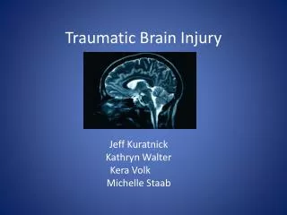
Traumatic Brain Injury. The effects of trauma to the skull and brain, during closed head injury depend upon a number of factors: (a) the shape of the objects causing the trauma, (b) the force of the impact, (c) whether the head is in motion at the time of impact.

E N D
The effects of trauma to the skull and brain, during closed head injury depend upon a number of factors: (a) the shape of the objects causing the trauma, (b) the force of the impact, (c) whether the head is in motion at the time of impact.
Traumatic brain injury (TBI) • An external mechanical force, • possibly leading to permanent or temporary impairments of : • cognitive, • physical, • psychosocial functions • with an associated diminished or altered state of consciousness.
High Risk Populations • Young people • Low-income individuals • Unmarried individuals • Men (2:1 vs. women) • Individuals with previous history of substance abuse • Individuals with previous TBI
Traumatic Brain Injury (TBI) • Brain injury results from: • Direct injury to brain tissue • External forces applied to outside of skull transmitted to the brain • Movement of brain inside skull
Thus the mechanisms of closed head injury are either due to • contactphenomena (usually resulting in focal lesions) or • acceleration/deceleration phenomena (principally causing diffuse axonal injury (DAI). • It is important to realize that severe brain damage can occur in the absence of easilyvisible external injury and vice versa, and that focal and diffuse lesions may occur in isolation or be combined.
HEAD INJURIES / BRAIN INJURIES CONCUSSION DIFFUSE AXONAL INJURY EPIDURAL SUBDURAL SUBARACHNOID PARENCHYMAL COUP/CONTRE COUP FOCAL/MULTIFOCAL INTRAVENTRICULAR
CONTACT PHENOMENA &/OR ACC/DEC INJURY PRIMARY DAMAGE EPIDURAL SUBDURAL SUBARACHNOID PARENCHYMAL COUP/CONTRE COUP FOCAL/MULTIFOCAL INTRAVENTRICULAR FOCAL DIFFUSE HEMATOMAS FRACTURES CONTUSIONS LACERATIONS HEMORRHAGES DIFFUSE AXONAL INJURY (DIFFUSE BRAIN INJURY)
Concussion • Concussion is caused by deformity of the deep structures of the brain, leading to widespread neurologic dysfunction that can result in impaired consciousness or coma. • Concussion is considered a mild form of diffuse axonal injury.
Diffuse axonal injury • Diffuse axonal injury is characterized by extensive generalized damage to the white matter of the brain. • Strains during high-speed acceleration/deceleration produced in lateral motions of the head may cause the injuries. • Diffuse axonal injury also could occur as a result of ischemia.
Diffuse axonal injury, in its mildest form is probably responsible for concussion, but if severe is associated with prolonged unconsciousness from the moment of injury. • The mechanism is one of widespread tearing of axons due to shearing forces during acceleration/deceleration of brain. • While focal injuries such as contusions, lacerations and intracranial hematomas, are usually readily apparent radiologically, DAI is more subtle.
CONSCIOUSNESS DISTURBANCE IN DIFFUSE BRAIN INJURY Mild concussion : LOC - transient Cerebral concussion : LOC <6hrs Mild DAI : LOC 6-24 hrs Moderate DAI : >24 hrs Severe DAI : days-weeks LOC:Loss of Consciousness DAI: Diffuse axonal injury
PATHOMORPHOLOGY OF DAI • Spheroids appear when coma exceeds 6 hrs • Only detectable at LM level after • 24 hours • Corpus callosum & brainstem most • commonly affected • Evanescent - days to weeks INVISIBLE ON CT/MR SCAN
Morphology of DAI Axonal spheroids
A.Dural associated hematomas Epidural Hematoma Subdural Hematoma
Epidural Hematoma • Normally the dura is fused with the periosteum on the internal surface of the skull. Dural arteries, most importantly the middle meningeal artery, are vulnerable to injury, particularly with temporal skull fractures in which the fracture lines cross the course of the vessel. In children, in whom the skull is deformable, a temporary displacement of the skull bones leading to laceration of a vessel can occur in the absence of a skull fracture.
Epidural Hematoma • The extravasation of blood under arterial pressure can cause the dura to separate from the inner surface of the skull . • The expanding hematoma has a smooth inner contour that compresses the brain surface. • When blood accumulates slowly patients may be lucid for several hours before the onset of neurologic signs. • An epidural hematoma may expand rapidly and is a neurosurgical emergency requiring prompt drainage.
Epidural Hematoma • More often, a tear in the middle meningeal artery causes this type of hematoma. • When hematoma occurs from laceration of an artery, blood collection can cause rapid neurologic deterioration.
Two-thirds of epidural hematomas are in the temporo-parietal region and they usually have a biconvex or lentiform configuration. • Epidurals are limited by the firmer attachment of the dura at the suture margins, but they may cross the midline.
Subdural hematoma • Subdural hematoma tends to occur in patients with injuries to the dural (cortical) veins or pial artery in severe TBI. • The associated mortality rate is high, approximately 60-80%. • They occur most frequently over the convexities, covering the entire hemisphere and are manifested clinically by fluctuating levels of consciousness.
Subdural hematomas, both acute and chronic, are most often caused by bleeding from torn bridging dural veins. • Subdural hematomas are less frequently associated with skull fractures, but more frequently associated with parenchymal brain damage. • The subdural space is a more freely communicating space and the hematomas form a crescentic shaped layer over the brain surface.
Instead, they extend along the dura of the falx into the interhemispheric fissure and onto the tentorium, which epidurals cannot do. • Both epidural and subdural hemorrhages occur within the confined space of the bony calvarium and compress the adjacent brain, often requiring emergency evacuation.
In alcoholic or elderly patients with cerebral atrophy, SDH's are often bilateral and may occur after insignificant or even unnoticed trauma.
bridging veins are more susceptible to tearing when there is cortical atrophy
Chronic subdural hematomas are usually related to a slower venous bleed without accompanying cerebral parenchymal injury. • These collections are more often biconvex, rather than the crescentic shape of acute subdural hematomas.
Subdural hygromas or effusions consist of collections of CSF in the subdural spaces, • presumably due to a traumatic arachnoid tear or they may also develop following ventricular shunting. • They may accumulate slowly during the first few days following head trauma, especially in the pediatric population.
Contusions &Lacerations • Contusions are focal areas of cortical hemorrhagic necrosis occurring at the crest of a gyrus and are said to be the hallmark of closed head injury; they may occur : • at the site of impact (coup lesions), or • at a point opposite (contrecoup lesions).
Contusion/laceration of the brain may be accompanied by pure intracerebral hematoma, often multiple, mainly involving temporal and frontal regions. • A distinction is made between contusions and lacerations: • Contusions (contusio cerebri): the pia is intact, • Lacerations(laceratio cerebri) : the pia-arachnoid and underlying brain tissue are torn.
Coup-Contrecoup Contusions • A combination of vascular and tissue damage leads to cerebral contusion. • Coup contusions occur at the area of direct impact to the skull and occur because of the creation of negative pressure when the skull, distorted at the site of impact, returns to its normal shape. • Contrecoup contusions are similar to coup contusions but are located opposite the site of direct impact.
Most of the energy of impact from a small hard object tends to dissipate at the impact site, leading to a coup contusion. • On the contrary, impact from a larger object causes less injury at the impact site since energy is dissipated at the beginning or end of the head motion, leading to a contrecoup contusion.
Traumatic intracerebral / intraventricular hemorrhages • Probably they result from tearing of the vessels themselves by mechanical forces.
VASCULAR INJURIES • Traumatic carotid cavernous fistulas can result from an injury to the carotid artery at the level of the cavernous sinus. • This causes a high flow arterial venous shunt and often results in painful unilateral pulsating exopthalmos as a result of increased flow in a retrograde direction in the superior opthalmic vein(v. ophthalmica superior). • This can lead to visual loss.
Traumatic dissections of the carotid or vertebral arteries in the neck can cause stroke. • This can be due to strangulation or whiplash mechanisms which may tear the intima. • Typically the carotid is dissected usually just proximal to entering the petrous bone at about the C1-C2 level.
Post-traumatic pseudoaneurysms can occur as a result of vascular injuries intracranially. • Vascular spasm can occur as a result of subarachnoid hemorrhage.
CEREBROVASCULAR DISEASE (CVD) CVD REFERS TO ANY ABNORMALITY OF THE BRAIN RESULTING FROM A PATHOLOGICAL PROCESS INVOLVING BLOOD VESSELS, CAUSING INADEQUATE OXYGENATION OF BRAIN TISSUE
CLASSIFICATION • HYPOXIC ENCEPHALOPATHY • an overall (GLOBAL) reduction in blood flow to the brain • STROKE (focal lesion) • sudden onset • rapid evolution • focal neurologic deficit
STROKE The typical clinical features include a sudden onset and rapid evolution (minutes/hours) of a focal neurological deficit (indicative of focal damage to the brain).
ISCHEMIC STROKE HEMORRHAGIC STROKE STROKE: CLASSIFICATION or INTRACEREBRAL BLEEDING or BRAIN INFARCTION THE CLINICIAN MUST MAKE THIS DISTINCTION IN ORDER TO TREAT CT/MR SCAN ALLOWS DISTINCTION
It is important to draw a distinction between ischemic stroke, • which refers to a focal cessation of blood supply due to vascular occlusion, resulting innecrosisof tissue (cerebral infarction) • and hemorrhagic stroke, • a term applied to rupture of a cerebral blood vessel resulting in spontaneous intracranial hemorrhage. In both instances, subsequent secondary events such as brain swelling and shifts (herniation) result in progressive coma and are the cause of death.
I. Ischemic stroke (CEREBRAL INFARCTION) • Although there are many variations regarding the evolution, site, size and shape of cerebral infarcts, certain patterns emerge based on an interplay of hemodynamic & etiological factors: • (a) the site of the occlusion (eg. distal/proximal/end artery), in relation to the presence and efficacy of a collateral blood supply (eg. anatomical variations, diseased and collateral vessels). • (b) type of occlusion - in general, thrombosis leading to gradual occlusion will allow adaptation of anastomoses, while emboli produce sudden occlusion.