Download

1 / 93
1.18k likes | 2.45k Vues
Traumatic Brain Injury. The First Affiliated Hospital, Medical College, Zhejiang University Neurosurgical Department Ying Tong. Epidemiology. Traumatic brain injury (TBI) continues to be an enormous public health problem. 500,000 to well over 1 million cases of head injury every year.

E N D
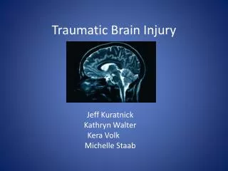
Traumatic Brain Injury The First Affiliated Hospital, Medical College, Zhejiang University Neurosurgical Department Ying Tong
Epidemiology • Traumatic brain injury (TBI) continues to be an enormous public health problem. • 500,000 to well over 1 million cases of head injury every year. • mild(80%), moderate(10%) and severe(10%) injuries. • Head injury results in half of the 150,000 trauma deaths. • 5.3 million people are living with brain injury–related disabilities.
Pathophysiology • Primary injury occurs at the moment of impact. Primary injury includes bone fracture, concussion, contusion, hematoma, traumatic subarachnoid hemorrhage, and diffuse axonal injury. • Secondary injury to the brain occurs as a result of decreased oxygen delivery to the brain, which in turn sets off a cascade of events that causes even more damage than the initial injury.
Pathophysiology • Primary injury • Concussion • Contusion • Intracranial hemorrhage: epidural, subdural, subarachnoid, and intraparenchymal or intracerebral • traumatic subarachnoid hemorrhage
Pathophysiology • Primary injury • Diffuse axonal injury(DAI) is a rotational acceleration-deceleration injury to the white-matter pathways of the brain resulting in a functional or anatomic disruption of these pathways and is cited as the cause of loss of consciousness in patients without mass lesions.
Pathophysiology • Secondary injury : Effects of hypotension, hypoxia, and herniation with elevated ICP due to mass effect, as a result of decreased oxygen delivery to the brain, which in turn sets off a cascade of events(mediators of inflammation, excitotoxicity, calcium influx, and Na+,K+-ATPase dysfunction ) leads to neuronal cell dysfunction and death.
Pathophysiology • Secondary intracranial insults to the brain • Hemorrhage • Ischemia • Edema • Raised intracranial pressure (ICP) • Vasospasm • Infection • Epilepsy • Hydrocephalus
Pathophysiology • Secondary systemic insults to the brain • Hypoxia • Hypercapnia • Hyperglycemia • Hypotension • Severe hypocapnia • Fever • Anemia • Hyponatremia
Mechanisms of brain injury Physical mechanisms of brain injury • Impact loading - Collision of the head with a solid object at a tangible speed • Impulsive loading - Sudden motion without significant physical contact • Static or quasistatic loading - Loading in which the effect of speed of occurrence may not be significant
Mechanisms of brain injury Basic types of tissue deformation • Compressive - Tissue compression • Tensile - Tissue stretching • Shear - Tissue distortion produced when tissue slides over other tissue
Mechanisms of brain injury • Coup injuries are caused by direct transmission of impact energy through the skull into the underlying brain and occur directly below the site of injury because of the creation of negative pressure when the skull, distorted at the site of impact, return to its normal shape.
Mechanisms of brain injury • Contrecoup injuries are similar to coup contusions but are located opposite the site of direct impact. Contrecoup injuries are caused by rotational shear and other indirect forces that occur contralateral to the primary injury. Rotational force causes the basal frontal and temporal cortices to impact or sweep across rigid aspects of the skull, the sphenoid wing, and petrous ridges. They are commonly seen in the frontal and temporal lobes.
Classification • By mechanism • Closed: High velocity (auto accidents); Low velocity (falls, assault) • Penetrating: Gunshot wounds; Other open injuries • By severity • Mild: GCS 13-15; Moderate: GCS 9-12; Severe: GCS 8 or less, comatose
Classification • By morphology • Skull fractures: Vault ---Linear or stellate ---Depressed or nondepressed Basilar ---With/without CSF leak --- With/without nerve VII palsy • Intracranial lesions: Focal --- Epidural --- Subdural --- Intracerebral Diffuse --- Mild concussion --- Classic concussion --- Diffuse axonal injury
Prehospital and Emergency Department Management • Cardiopulmonary stabilization :ABCs ; avoid hypotension and hypoxia; systolic blood pressure> 90 mm Hg , PaO2 > 60 mm Hg. • Medical history: AMPLE • General examination • Neurologic examination: The Glasgow Coma Scale (GCS)and the pupils.
Initial neurologic examination • Glasgow Coma Scale • Pupillary response to light • Eye movement • Oculocephalic (doll’s eye) • Oculovestibular (caloric) • Motor power • Gross sensory examination
Neurologic examination: the pupils • Pupillary size and reactivity are also essential components of the initial neurologic examination. • The presence of an unresponsive pupil, particularly a dilated pupil, is significantly correlated with a poor outcome and therefore an important part of the examination.
Interpretation of pupillary findings in head injury patients
Diagnostic procedure • CT scanning:greater efficacy ; emergency CT scan ; assess the presence or absence of fracture, epidural and subdural hematomas, intracerebral hematomas and contusions, shift of the midline structures, and the appearance of the basal and perimesencephalic cisterns. • plain skull films .
CT scan of the head in a patient with a closed head injury. Severe compression of mesencephalic cisterns is seen, indicating midbrain compression (arrowheads).
CT scans of patient with closed head injury. This patient has a right temporal epidural hematoma (arrows). The mesencephalic cisterns are patent in the top left, indicating a lack of brain stem compression despite mass (arrowheads).
CT scans of patient with closed head injury.This patient has suffered an acute left subdural hematoma (arrowheads) with midline shift (arrows).
Intracranial Pressure • Indication:all patients with a GCS of 8 or less have ICP monitors placed. • Types: IVCs and sites other than the ventricles ( brain parenchyma, subarachnoid or subdural or epidural space).
Nonoperative Management • Prompt physiologic resuscitation: restoration of blood pressure, oxygenation, and ventilation. • Control increased ICP: The goal of treatment is to try to keep the ICP less than 20mm Hg and to maintain cerebral perfusion pressure at or above 70 mm Hg.
Control increased ICP • The head of the bed is elevated to 30 degrees with the head placed in a neutral position. • CSF drainage: external ventricular drain, or ventriculostomy. • Mannitol and other diuretic agents: Mannitol --- IV bolus of 0.25 to 1 g/kg body weight every 4 to 6 hours. Other diuretics : frusemide .---maintain euvolemia
Control increased ICP • Hyperventilation: PaCO2 of 30 to 35 mm Hg . lowering PaCO2 ----- vasoconstriction -----decreases intracranial blood volume----- a decrease in ICP • Mild hypothermia • Second-tier therapeutic interventions: hypertonic saline, high-dose barbiturate therapy, decompressive craniectomy.
Other Nonoperative Management • nutrition : nonparalyzed patient , 140% of their resting metabolism expenditure; paralyzed patient , 100%; • Steroids:not recommend; • Anticonvulsants: prevent early post-traumatic seizures .
Operative Management • Indication: an intracranial mass lesion and deficits thought to be related to that lesion. [Space-occupying hematomas , the amount of midline shift, the location of the clot and the patient’s ICP ] • In general, any clot or contusion greater than 30 cc is thought to be operable.
Outcome • Outcome has improved since the mid 1970s . • Mortality rates have decreased from approximately 50% to less than 30% .
Algorithm for management of mild head injury. *Risk factors include: ambiguous accident history, continued post-traumatic amnesia, retrograde amnesia > 30 min, clinical signs of skull fracture, headache, vomiting, focal neurologic deficit, seizure, age < 2 years and > 60 years, coagulation disorder, or high-energy (speed) accident.
Introduction • Classification : by pattern (linear, comminuted, depressed), by anatomic location (vault convexity, base), and by skin integrity (open, closed). • The pattern of a skull fracture: the force of impact and the ratio of the impact force to the impact area.
Linear skull fractures • A linear skull fracture is a single fracture line that goes through the entire thickness of the skull. • Diagnosis: CT scan, plain skull radiographs. • Management: closed linear fractures require no stabilization or exploration when there is no evidence of epidural hematoma or underlying dural or cortical injury. Open linear fractures are debrided.
Comminuted fractures • A comminuted fracture occurs when multiple linear fractures radiate from the point of impact. • Diagnosis: skull radiographs and CT. • Management :1) If skin is closed and no depression of bone fragments greater than the thickness of the skull, management is as for linear skull fractures. 2)With underlying intracranial pathology,surgery. 3)Open and free bone fragments are present, cleansing or debridement.