Download
1 / 30
300 likes | 524 Vues
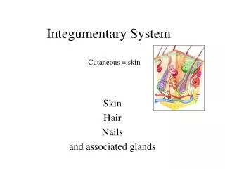
Integumentary System (Skin). Skin Functions. Protects deeper tissues from: Mechanical damage ( bumps ) Chemical damage ( acids and bases ) Bacterial damage Ultraviolet radiation ( sunlight ) Thermal damage ( heat or cold ) Desiccation ( drying out )

E N D
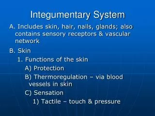
Skin Functions • Protects deeper tissues from: • Mechanical damage (bumps) • Chemical damage (acids and bases) • Bacterial damage • Ultraviolet radiation (sunlight) • Thermal damage (heat or cold) • Desiccation (drying out) • Aids in body heat loss or heat retention as controlled by the nervous system • Aids in excretion of urea and uric acid • Synthesizes vitamin D
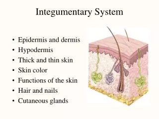
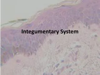
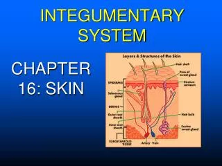
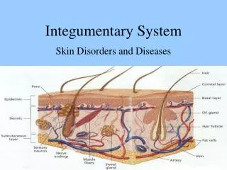
Skin Structure • Epidermis—outer layer • Stratified squamous epithelium • Can become keratinized (hardened by keratin) to prevent water loss • Avascular (no blood supply of its own) • Most cells are keratinocytes (cells that produce keratin – a fibrous protein that makes the epidermis tough) • Dermis • Dense connective tissue • Subcutaneous tissue (hypodermis) is deep to dermis • Not technically part of the skin • Anchors skin to underlying organs • Composed mostly of adipose tissue
Layers of the Epidermis • Can contain up to 5 of these layers (strata) • Stratum basale (stratum germinativum) • Deepest layer of epidermis • Lies next to dermis • Wavy borderline with the dermis anchors the two together • Cells undergoing mitosis (cell division) • Daughter cells are pushed upward to become the more superficial layers • Stratum spinosum • Stratum granulosum Cells become flatter and more full of keratin
Layers of the Epidermis (cont’d) • Stratum lucidum • Formed from dead cells of the deeper strata • Occurs only in thick, hairless skin of the palms of hands and soles of feet • Stratum corneum • Outermost layer of epidermis • Shingle-like dead cells are filled with keratin (protective protein prevents water loss from skin)
Summary of layers from deepest to most superficial • Stratum basale • Stratum spinosum • Stratum granulosum • Stratum lucidum (thick, hairless skin only) • Stratum corneum
Melanin • Pigment (melanin) produced by melanocytes • Melanocytes are mostly in the stratum basale • Color is yellow to brown to black • Amount of melanin produced depends upon genetics and exposure to sunlight
Dermis • Two layers • Papillary layer (upper dermal region) • Projections called dermal papillae • Some contain capillary loops • Others house pain receptors and touch receptors • Reticular layer (deepest skin layer) • Blood vessels • Sweat and oil glands • Deep pressure receptors
Dermis (cont’d) • Overall dermis structure • Collagen and elastic fibers located throughout the dermis • Collagen fibers give skin its toughness • Elastic fibers give skin elasticity • Blood vessels play a role in body temperature regulation
Normal Skin Color Determinants • Melanin • Yellow, brown, or black pigments • Carotene • Orange-yellow pigment from some vegetables • Hemoglobin • Red coloring from blood cells in dermal capillaries • Oxygen content determines the extent of red coloring
Alterations in Skin Color • Redness (erythema)—due to embarrassment, inflammation, hypertension, fever, allergy, or conditions like rosacea • Pallor (blanching)—due to emotional stress such as fear, anemia, low blood pressure, impaired blood flow to an area • Jaundice (yellowing)—liver disorder • Bruises—hematomas
Skin Appendages • Sebaceous (Oil) glands • Sudoriferous (Sweat) glands • Hair • Hair follicles • Nails
Sebaceous (oil) glands • Produce oil (sebum) • Lubricant for skin • Prevents brittle hair • Kills bacteria • Most have ducts that empty into hair follicles; others open directly onto skin surface • Glands are activated at puberty
Sudoriferous (Sweat) glands • Produce sweat • Widely distributed in skin • Two types of sudoriferous glands • Eccrine • Open via duct to pore on skin surface • Produce sweat (clear) • Apocrine(Begin to function at puberty) • Ducts empty into hair follicles • Release sweat that also contains fatty acids and proteins (milky/yellowish color)
Sweat and its Function • Composition • Mostly water • Salts and vitamin C • Some metabolic waste • Fatty acids and proteins (apocrine only) • Function • Helps dissipate excess heat • Excretes waste products • Acidic nature inhibits bacteria growth • Odor is from associated bacteria
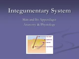
Hair • Produced by hair follicle • Consists of hard keratinized epithelial cells • Melanocytes provide pigment for hair color • Hair grows in the matrix of the hair bulb in the stratum basale • Hair anatomy • Central medulla • Cortex surrounds medulla • Cuticle on outside of cortex • Most heavily keratinized
Associated Hair Structures • Hair follicle • Dermal and epidermal sheath around hair root • Arrector pili muscle • Smooth muscle • Pulls hair upright when cold or frightened • Sebaceous gland • Sudoriferous gland
Nails • Scale-like modifications of the epidermis • Heavily keratinized • Stratum basale extends beneath the nail bed • Responsible for growth • Lack of pigment makes them colorless
Nail Structures • Free edge • Body: visible attached portion • Root: embedded in skin • Cuticle: proximal nail fold that projects onto the nail body
Burns • Tissue damage and cell death caused by heat, electricity, UV radiation, or chemicals • Associated dangers • Dehydration • Electrolyte imbalance • Circulatory shock
Rule of Nines • Way to determine the extent of burns • Body is divided into 11 areas for quick estimation • Each area represents about 9 percent of total body surface area
Severity of Burns • First-degree burns • Only epidermis is damaged • Skin is red and swollen • Second-degree burns • Epidermis and upper dermisare damaged • Skin is red with blisters • Third-degree burns • Destroys entire skin layer; burned area is painless • Burn is gray-white or black
Critical Burns • Burns are considered critical if • Over 25 percent of body has second-degree burns • Over 10 percent of the body has third-degree burns • There are third-degree burns of the face, hands, or feet
Skin Homeostatic Imbalances Infections
Infections • Athlete’s foot (tineapedis) • Caused by fungalinfection • Boils and carbuncles • Caused by bacterialinfection • Cold sores • Caused by virus
Infections & Allergies • Contact dermatitis • Exposures cause allergic reaction • Impetigo • Caused by bacterialinfection • Psoriasis • Cause is unknown • Triggered by trauma, infection, stress