Encoding of visual stimulus
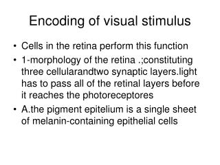
Encoding of visual stimulus. Cells in the retina perform this function 1-morphology of the retina .;constituting three cellularandtwo synaptic layers.light has to pass all of the retinal layers before it reaches the photoreceptores

Encoding of visual stimulus
E N D
Presentation Transcript
Encoding of visual stimulus • Cells in the retina perform this function • 1-morphology of the retina .;constituting three cellularandtwo synaptic layers.light has to pass all of the retinal layers before it reaches the photoreceptores • A.the pigment epitelium is a single sheet of melanin-containing epithelial cells
Pigment epithelium functions • Light absorption • Phagocytosis • Vitamin A (retinol storage) • Clinical significance:retinal detachment can lead to blindness
Retinal cellular layers • Outer nuclear layer (OLN):Nuclei of photo receptors • Inner L N :Nuclei of bipolar and horizontal • Ganglion cell layer
Synaptic layers • Outer plexiform layer(OPL) • Inter Plexiform Layer(IPL)
Receptive fields • Each ganglion cell collects information from a group of receptors called its receptive field • A.circular receptive field;in fovea 10 um • Near the periphery 1 mm in diameter • 1-The cones in the center of RF convey directly to bipolar cells • 2- 1-The cones in the periphery of RF convey indirectly through horizontal cells
RF properties • 1-center surrond antagonistic region • A-on center receptive field • B-off center receptive field • Illuminating surrond (on center RF) • Illuminating center and surrond ,no effect on ganglion cell firing
Visual pathway • From ganglion cells to to visual cortex within LGN • Optic nerve-optic chiasma-right half of each retina to right LGN • Temporal (uncrossed fiber go to 2,3 5 • Crossed go to 1,4,6
Visual perception The receptive field organization of the retina and cortex are used to encode information about intensity ,contrast ,form ,color and depth
1- light intensity Light intensity is encoded by the firing rate of ganglionn cells a-at a very low light levels,only rods are active b-at the high light levels ,only cones are involved in the stimulation of ganglion cells
2-Contrast Contrast is encoded when one ganglion cell is stimulated and its neighbor is inhibited A-neighboring cells respond in opposite ways at the border between light and dark areas of the image
Contrast cot. 1-in the lighted portions of the border between light and dark ,the center of the receptive field is illuminated ,while its surround is not .therefore ,the on-center ganglion cell activity is increased 2- in the darkened portions ,the surrond of the receptive field is illuminated ,ehile its center is not. therefore ,the on-center ganglion cell activity is decresed
Contrast cot. B-when both the center and surrond of thereceptive field are totally in the illuminated or darkened portions of the image ,no change in ganglion cell activity occurs.
Form Information about form is decoded by cell within the primary visual cortex (simple cells) A-simple cells have center-surround receptive field .(rectangularly organized ) B-ganglion cells from each area of the retina project ,via the LGN,to a cortical cells organized into columns.each column contains simple cells oriented in a particular direction.
Form cont. • 1-the orientation of the simple cells in a small column(50um in wide )columnar region of the cortex is the same . • 2-adjacent columns have orientations that are approximately 10 degree apart;therefore lines of any angle at any point on the retina are decoded by specific Column of cells in the cortex
Depth Information about depth is provided by cells within areas 18,19 and are organized into hypercolumns. a.ocular-dominant cells are binocular (they recive input from both eyes) and fuse the images in the hypercolumns . b.The amount of disparity between the images percived by each eye is used to assign depth to the image .Depth perception (stereopsis)is possible because each eye percives a slightly different image.
Color • The cells responsible for color perception are organized into oblong regions called blobs .the blobs are located within the hypercolumn of cells that decode form and depth . • A.ganglion cells relaying information about color to LGN respond oppsitely to red and green light ;therefore,they are called color-opponent cells.
Color cont. • Cortical cells interpret the information recived from the LGN to assign color to each region of the image formed on the retina .For example ,if green center and red center cortical cells are equally excited,the color assigned the region of the cortex is yellow.
Color blindness • Color blindness results from the absence of the gene that codes for either the red ,green,or blue opsin. • Red-green color blindness results from the inability to synthesize either red or green pigment .this genetic defet is sex-linked .approximately 9% of the male population hassome sort of red or green color deficit. • Blue color blindness is very rare.
Structure of the ear Three divisions ,external ,middle ,inner External Pina:helps identify sound source external auditory canal:
Middle ear:Air filled • tympanic membrane (ear drum) , • three small bone (autitory ossicles) • Malleus ,incus, • stapes