Download
1 / 21
220 likes | 472 Vues
Identification of Laser Dissected Cotton Chromosomes using PCR-based markers. TaShundra J. Bryant. Outline of talk:. Introduction - Cotton - Markers Materials & Methods - Laser Capture Micro-dissection (LCM) microscope. - Whole Genome Amplification (WGA)

E N D
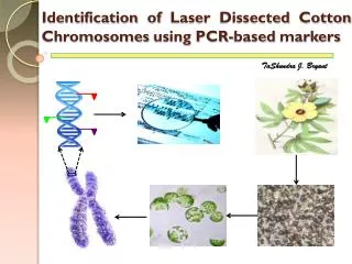
Identification of Laser Dissected Cotton Chromosomes using PCR-based markers TaShundra J. Bryant
Outline of talk: • Introduction • - Cotton • - Markers • Materials & Methods • - Laser Capture Micro-dissection (LCM) microscope. • - Whole Genome Amplification (WGA) • - Polymerase Chain Reaction (PCR) • - Poly-Acrylamide Gel Electrophoresis (PAGE) • Results • Discussion • Conclusion
Introduction: • Cotton(Gossypium spp.) • Fiber & oil. • Genus cotton (50 sps) • - Diploid species (2n = 2x = 26) ~ 45 species. • {differentiated cytogenetically into A-G & K groups}. • - Allotetraploid (2n = 4x = 52) ~ 5 species. • G. hirsutum ~ 95% world cotton production. • (upland cotton) ~ Genome size 2,500Mb (30k genes).
Introduction: (continued…) • Molecular Marker • “- a readily detectable sequence of DNA or protein whose inheritance can be monitored”. • Key characteristics: • - polymorphic, • - reproducible, • - co-dominant and • - fast and inexpensive to detect. • Types: Bio-chemical Non-PCR based PCR based
Objectives: • to dissect the cotton chromosomes using LCM. • to isolate the DNA and to amplify the product using Whole Genome Amplification method. • to identify the chromosomes using chromosome specific PCR based markers.
Outline of work: Germinating the cotton seeds Isolating the protoplasts from germinated seedlings Spreading the protoplast on PET membrane slides Picking the individual chromosomes using LCM Isolation of chromosomes Amplification of chromosomes using WGA
Amplified WGA3 product Selecting the SSR makers for screening PCR amplification using selected markers Running the PCR product on PAGE gel PAGE gel Analysis Identification of chromosome
Materials & Methods: • The genetic standard, G. hirsutum TM-1 (Texas Marker-1) was used for the preparation of protoplasts. • Protoplasts were isolated and spread on polyethylene tetraphthalate (PET) membrane slide. • Individual chromosomes were isolated using Laser Capture Micro-dissection microscope (Veritas). • 25 chromosome samples, B2, B3, B4, B8, B9, B11, B12, B13, A2, A4, A5, A8, A9, A10, A13, A14, A17, A18, A19, A20, A21, A27, A28, A32, A33, TM-1 and -Ve were used for further analysis.
Chromosome specific markers: BNL3279 BNL1066 BNL836 BNL1231 BNL625 BNL3411 BNL3431 MUSS123 CAPS & EST-SSRs BNL2572 BNL4049 BNL530 BNL4047 NAU1267 Fig.1a. Chromosome 4 map Fig.1a. Chromosome 11 map
LCM: (UV laser microdissection) PET membrane slide UV laser photovolatilizes cells Laser ablation of unwanted cells Cells and membrane are cut from tissue
PCR: Denaturation (940C) Annealing (570C) Extension (720C)
PAGE: • 6% PAGE gels were used • acrylamide:bisacrylamide (29:1) • PCR product was runned at 250V for 2:30 min’s • Visualized under UV light after ethidium bromide staining. • Gel images were saved and scoring was carried out manually. Vertical gel electrophoresis systems (C.B.S. Scientific Company, inc.)
Results: Fig.1a. Locating the chromosome using LCM Fig.1b. Dissecting the chromosome using LCM
Results: (continued…) Marker Marker 300 bp 300 bp B4 B9 A2 A9 B2 A5 B12 A13 A17 A19 A21 A28 A33 200 bp 200 bp A27 B13 B3 B8 B11 A10 A14 A18 A20 A32 A4 A8 TM-1 -VE 100 bp 100 bp Fig.2. PAGE gel Analysis for BNL-530 marker
Discussion: • Of 13 markers used, 7 markers were positive with selected samples while 6 markers were negative to all samples
Discussion: (Continued…) • Using chromosome specific markers, the samples A33 and A27 were potentially identified as a chromosome 11 and chromosome 4 respectively. • Samples B12, A9, A13 and A17 were neutral with selected markers, indicating that they were potential candidates for other chromosome specific markers.
Discussion: (Continued…) • Possible reasons for no amplification: • - due to genotypic differences of the parents or • - due to lack of proper PCR conditions • - due to error in assigning the marker to mapped loci.
Conclusions: • Use of chromosome-specific markers will aid in sequencing of individual chromosomes. • It also aids in analyzing the genetic diversity among cultivated and wild cotton species. • Integrating the available polymorphic markers such as CAPS, EST-SSRs etc and linkage groups help in developing comprehensive cotton genetic map to assist the cotton breeders.
Acknowledgements: • Dr. Govind Sharma • Dr. Ramesh Kantety • Dr. Ramesh Buyyarapu • Sripathi Venkat & • All members of CMB group