Download
1 / 57
720 likes | 2.02k Vues
Cardiac Physiology. Cardiac Physiology - Anatomy Review. Circulatory System. Three basic components Heart Serves as pump that establishes the pressure gradient needed for blood to flow to tissues Blood vessels

E N D
Circulatory System • Three basic components • Heart • Serves as pump that establishes the pressure gradient needed for blood to flow to tissues • Blood vessels • Passageways through which blood is distributed from heart to all parts of body and back to heart • Blood • Transport medium within which materials being transported are dissolved or suspended
Circulatory System • Pulmonary circulation • Closed loop of vessels carrying blood between heart and lungs • Systemic circulation • Circuit of vessels carrying blood between heart and other body systems
Functions of the Heart • Generating blood pressure • Routing blood • Heart separates pulmonary and systemic circulations • Ensuring one-way blood flow • Regulating blood supply • Changes in contraction rate and force match blood delivery to changing metabolic needs
Electrical Activity of Heart • Heart beats rhythmically as result of action potentials it generates by itself (autorhythmicity) • Two specialized types of cardiac muscle cells • Contractile cells • 99% of cardiac muscle cells • Do mechanical work of pumping • Normally do not initiate own action potentials • Autorhythmic cells • Do not contract • Specialized for initiating and conducting action potentials responsible for contraction of working cells
Cardiac Muscle Cells • Myocardial Autorhythmic Cells • Membrane potential “never rests” pacemaker potential. • Myocardial Contractile Cells • Have a different looking action potential due to calcium channels. • Cardiac cell histology • Intercalated discs allow branching of the myocardium • Gap Junctions (instead of synapses) fast Cell to cell signals • Many mitochondria • Large T tubes
Intrinsic Cardiac Conduction System Approximately 1% of cardiac muscle cells are autorhythmic rather than contractile 70-80/min 40-60/min 20-40/min
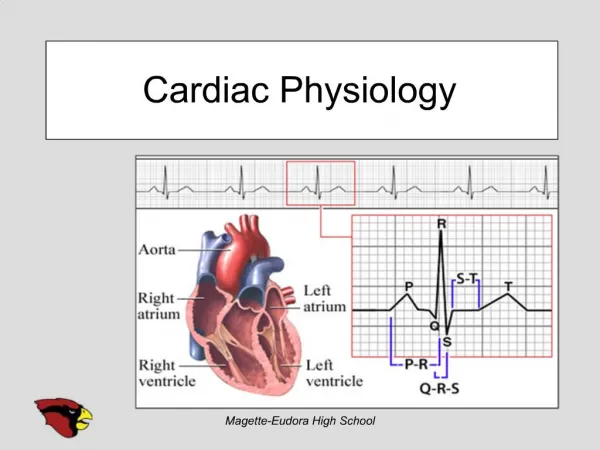
Electrocardiogram (ECG) • Record of overall spread of electrical activity through heart • Represents • Recording part of electrical activity induced in body fluids by cardiac impulse that reaches body surface • Not direct recording of actual electrical activity of heart • Recording of overall spread of activity throughout heart during depolarization and repolarization • Not a recording of a single action potential in a single cell at a single point in time • Comparisons in voltage detected by electrodes at two different points on body surface, not the actual potential • Does not record potential at all when ventricular muscle is either completely depolarized or completely repolarized
Asystole Regularity: Rate: P Waves: PRI: QRS: Straight line indicates absence of electrical activity Bad JUJU, Dude
Heart Attack • Chest Discomfort • Shortness of Breath • Nausea • Vomiting • Sweating • Dizziness • Palpitations • Syncope • Collapse/Sudden Death
Electrical Conduction • SA node - 75 bpm • Sets the pace of the heartbeat • AV node - 50 bpm • Delays the transmission of action potentials • Purkinje fibers - 30 bpm • Can act as pacemakers under some conditions
Intrinsic Conduction System • Autorhythmic cells: • Initiate action potentials • Have “drifting” resting potentials called pacemaker potentials • Pacemaker potential - membrane slowly depolarizes “drifts” to threshold, initiates action potential, membrane repolarizes to -60 mV. • Use calcium influx (rather than sodium) for rising phase of the action potential
Pacemaker Potential • Decreased efflux of K+, membrane permeability decreases between APs, they slowly close at negative potentials • Constant influx of Na+, no voltage-gated Na + channels • Gradual depolarization because K+ builds up and Na+ flows inward • As depolarization proceeds Ca++ channels (Ca2+ T) open influx of Ca++ further depolarizes to threshold (-40mV) • At threshold sharp depolarization due to activation of Ca2+ L channels allow large influx of Ca++ • Falling phase at about +20 mV the Ca-L channels close, voltage-gated K channels open, repolarization due to normal K+ efflux • At -60mV K+ channels close
PX = Permeability to ion X PNa 1 +20 2 PK and PCa 0 -20 PK and PCa 3 0 -40 Membrane potential (mV) PNa -60 -80 4 4 -100 0 100 200 300 Time (msec) Phase Membrane channels 0 Na+ channels open 1 Na+ channels close 2 Ca2+ channels open; fast K+ channels close 3 Ca2+ channels close; slow K+ channels open 4 Resting potential AP of Contractile Cardiac cells • Rapid depolarization • Rapid, partial early repolarization, prolonged period of slow repolarization which is plateau phase • Rapid final repolarization phase
AP of Contractile Cardiac cells • Action potentials of cardiac contractile cells exhibit prolonged positive phase (plateau) accompanied by prolonged period of contraction • Ensures adequate ejection time • Plateau primarily due to activation of slow L-type Ca2+ channels
Why A Longer AP In Cardiac Contractile Fibers? • We don’t want Summation and tetanus in our myocardium. • Because long refractory period occurs in conjunction with prolonged plateau phase, summation and tetanus of cardiac muscle is impossible • Ensures alternate periods of contraction and relaxation which are essential for pumping blood
Excitation-Contraction Coupling in Cardiac Contractile Cells • Ca2+ entry through L-type channels in T tubules triggers larger release of Ca2+ from sarcoplasmic reticulum • Ca2+ induced Ca2+ release leads to cross-bridge cycling and contraction
Electrical Signal Flow - Conduction Pathway • Cardiac impulse originates at SA node • Action potential spreads throughout right and left atria • Impulse passes from atria into ventricles through AV node (only point of electrical contact between chambers) • Action potential briefly delayed at AV node (ensures atrial contraction precedes ventricular contraction to allow complete ventricular filling) • Impulse travels rapidly down interventricular septum by means of bundle of His • Impulse rapidly disperses throughout myocardium by means of Purkinje fibers • Rest of ventricular cells activated by cell-to-cell spread of impulse through gap junctions
1 1 SA node AV node 2 1 THE CONDUCTING SYSTEM OF THE HEART SA node depolarizes. 2 Electrical activity goes rapidly to AV node via internodal pathways. SA node 3 Internodal pathways 3 Depolarization spreads more slowly across atria. Conduction slows through AV node. AV node 4 Depolarization moves rapidly through ventricular conducting system to the apex of the heart. A-V bundle 4 Bundle branches Purkinje fibers Depolarization wave spreads upward from the apex. 5 5 Purple shading in steps 2–5 represents depolarization. Electrical Conduction in Heart • Atria contract as single unit followed after brief delay by a synchronized ventricular contraction
Electrocardiogram (ECG) • Different parts of ECG record can be correlated to specific cardiac events
P wave: atrial depolarization START P The end R PQ or PR segment: conduction through AV node and A-V bundle T P P QS Atria contract. T wave: ventricular Repolarization ELECTRICAL EVENTS OF THE CARDIAC CYCLE Repolarization R T P QS Q wave P Q ST segment R R wave P R Q S P R Ventricles contract. Q P S wave QS Heart Excitation Related to ECG
ECG Information Gained • (Non-invasive) • Heart Rate • Signal conduction • Heart tissue • Conditions
Cardiac Cycle - Filling of Heart Chambers • Heart is two pumps that work together, right and left half • Repetitive contraction (systole) and relaxation (diastole) of heart chambers • Blood moves through circulatory system from areas of higher to lower pressure. • Contraction of heart produces the pressure
Late diastole: both sets of chambers are relaxed and ventricles fill passively. 1 START Isovolumic ventricular relaxation: as ventricles relax, pressure in ventricles falls, blood flows back into cups of semilunar valves and snaps them closed. 5 Atrial systole: atrial contraction forces a small amount of additional blood into ventricles. 2 Isovolumic ventricular contraction: first phase of ventricular contraction pushes AV valves closed but does not create enough pressure to open semilunar valves. 3 Ventricular ejection: as ventricular pressure rises and exceeds pressure in the arteries, the semilunar valves open and blood is ejected. 4 Cardiac Cycle - Mechanical Events Figure 14-25: Mechanical events of the cardiac cycle
Heart Sounds • First heart sound or “lubb” • AV valves close and surrounding fluid vibrations at systole • Second heart sound or “dupp” • Results from closure of aortic and pulmonary semilunar valves at diastole, lasts longer
Cardiac Output (CO) and Reserve • CO is the amount of blood pumped by each ventricle in one minute • CO is the product of heart rate (HR) and stroke volume (SV) • HR is the number of heart beats per minute • SV is the amount of blood pumped out by a ventricle with each beat • Cardiac reserve is the difference between resting and maximal CO
Cardiac Output = Heart Rate X Stroke Volume • Around 5L : (70 beats/m 70 ml/beat = 4900 ml) • Rate: beats per minute • Volume: ml per beat • SV = EDV - ESV • Residual (about 50%)
Factors Affecting Cardiac Output • Cardiac Output = Heart Rate X Stroke Volume • Heart rate • Autonomic innervation • Hormones - Epinephrine (E), norepinephrine(NE), and thyroid hormone (T3) • Cardiac reflexes • Stroke volume • Starlings law • Venous return • Cardiac reflexes
Factors Influencing Cardiac Output • Intrinsic: results from normal functional characteristics of heart - contractility, HR, preload stretch • Extrinsic: involves neural and hormonal control – Autonomic Nervous system
Stroke Volume (SV) • Determined by extent of venous return and by sympathetic activity • Influenced by two types of controls • Intrinsic control • Extrinsic control • Both controls increase stroke volume by increasing strength of heart contraction
Stroke volume Strength of cardiac contraction End-diastolic volume Venous return Intrinsic Factors Affecting SV • Contractility – cardiac cell contractile force due to factors other than EDV • Preload – amount ventricles are stretched by contained blood - EDV • Venous return - skeletal, respiratory pumping • Afterload – back pressure exerted by blood in the large arteries leaving the heart
Frank-Starling Law • Preload, or degree of stretch, of cardiac muscle cells before they contract is the critical factor controlling stroke volume