Download
1 / 15
150 likes | 392 Vues
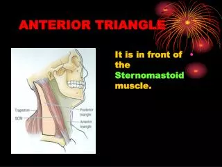
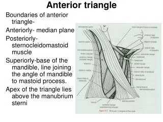
ANTERIOR TRIANGLE of the NECK. Borders. Superior: Mandible. Posterior: Sternocleidomastoid (anterior border). Anterior: Midline of the neck. Subdivisions. Muscular triangle. Submandibular triangle. Carotid triangle. Contents of Muscular Triangle. Digastric:

E N D
Borders • Superior: Mandible. • Posterior: Sternocleidomastoid (anterior border). • Anterior: Midline of the neck.
Subdivisions • Muscular triangle. • Submandibular triangle. • Carotid triangle.
Contents of Muscular Triangle • Digastric: Anterior and posterior bellies. Innervated by two different cranial nerves. • Stylohyoid. • Mylohyoid.
Contents of Muscular Triangle • Geniohyoid. • Sternohyoid. • Sternothyroid. • Thyrohyoid. • Omohyoid.
Carotid System • Common carotid. • Bifurcation: Blood monitors: Carotid sinus: Dilated terminal portion of common carotid. Baroreceptor. Carotid body: Chemoreceptor. Internal carotid: Has no branches in the neck. External carotid.
External Carotid Branches • Superior thyroid (anterior). • Ascending pharyngeal (medial). • Lingual (anterior): Crossed by hypoglossal nerve at its origin. • Facial (anterior). • Occipital (posterior). • Posterior auricular (posterior). • Superficial temporal (terminal). • Maxillary (terminal).
Characteristics and Branches of Cervical Plexus • From ventral rami of C1-4. • Hypoglossal nerve (CN XII). • Sensory branches: Great auricular (C2-3). Lesser occipital (C2(3)): Ventral ramus of C2. Greater occipital is dorsal ramus of C2. Transverse cervical (C2-3). Supraclavicular (C3-4).
Characteristics and Branches of Cervical Plexus • Motor branches: To prevertebral muscles: See syllabus. To levator scapulae muscle (C3-4). To scalene muscles. Ansa cervicalis: Supplies infrahyoid muscles. Except thyrohyoid. Phrenic nerve (C3,4,5).
Ansa Cervicalis • Nerve to geniohyoid: C1 via hypoglossal nerve. • Nerve to thyrohyoid. • Superior and inferior roots: Omohyoid. Sternothyroid. Sternohyoid.
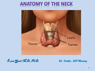
Deep Neck • Subclavian artery: Review from syllabus. • Scalene muscles: Review from syllabus. • Thyroid gland. • Trachea. • Esophagus. • Thoracic duct and right lymphatic duct.
Deep Neck • Branches of vagus nerve: Review from syllabus. • Sympathetic trunk: Ascends on anterior surfaces of longus colli and longus capitis muscles. Lies posterior to common and internal carotids. Lies deep to carotid sheath. Contains preganglionic sympathetic fibers from T1 and T2 and T2 and GVA fibers as well from same levels.
Sympathetic Trunk Ganglia • Superior cervical ganglion: Elongated structure on longus capitis muscle. At level of C1 and C2. Postganglionic sympathetic fibers from this ganglion supply all smooth muscles and glands of the head.
Sympathetic Trunk Ganglia • Middle cervical ganglion. • Inferior cervical ganglion. • Stellate ganglion: Fused inferior cervical ganglion and T1 ganglion.