Download
1 / 128
1.37k likes | 2.36k Vues
RHEUMATIC FEVER. Dr. Haripriya Jayakumar Senior resident. Introduction History Epidemiology Etiology Immunopathogenesis Pathology Clinical features Diagnosis Treatment Prevention. Introduction. Rheumatic fever is – inflammatory systemic disease

E N D
RHEUMATIC FEVER Dr. HaripriyaJayakumar Senior resident
Introduction • History • Epidemiology • Etiology • Immunopathogenesis • Pathology • Clinical features • Diagnosis • Treatment • Prevention
Introduction • Rheumatic fever is – inflammatory systemic disease triggered by Group A Streptococcus (non-suppurative complication) genetically predisposed individuals • Leading cause of acquired heart disease in children and young adult worldwide
WORLD WIDE INCIDENCE 1970-1990 Seckeler, Hoke: The worldwide epidemiology of acute rheumatic fever and rheumatic heart disease; Clinical Epidemiology, 2011
Names to be remembered.. Rheumatic fever described since the 17th century • Sydenham – related chorea with arthitis 1600s • Charles Wells (1812) – Association of rheumatism with carditis and subcutaneous nodules • Jean –BaptisteBouillaud (1836) – 1st publication on rheumatic fever (‘Father of rheumatic heart disease’) • Walter B Cheadle(1889)– 1st classic description of rheumatic fever.
Ludwig Aschoff(1904) - 1st description of pathology of rheumatic carditis. • Alvin F Coburn (1931) – 1st proved causal relationship between Beta hemolytic Strep and ARF • Rebecca Lancefield (1933) – Serogrouping of Streptococci • T Duckett Jones (1944) – 1st diagnostic criteria for RF. He presented his paper on the diagnosis of rheumatic fever in Chicago (AMA meeting)
The agent - Group A beta hemolytic streptococci Gram positive cocci chain / pair Non sporing, non motile Facultative anerobes Encapsulated
Lancefield serological classification On the basis of the C carbohydrate antigen on the cell wall Streptococci classified into many groups from A-K & H-V
The M protein: • major surface antigen • epitopes which cross react with myocardium, synovia, skin and brain • Alpha helical coiled-coil fibrillar protein– significant homology in structure & amino acid sequences to tropomyosin, myosin, keratin, vimentin & laminin.
Based on the conserved C repeat regions Class I & Class II GAS strains are named. • It is the Class I M-type of which belongs the strains 1,3,5,6,14,18,19 and 24 ---that have been associated with RF
GAS pharyngitis • Age group : 5-15 years • In epidemics, 3 % of untreated cases – developed RF • In endemic areas, 0.3% of untreated cases developed • Infection may be asymptomatic in 30-50% of cases of RF (approx one-third)
The environment • Overcrowding • Poverty • Poor nutrition • Poor hygiene • Poor access to health care • Rapid spread • Lack of primary prevention • Lack of secondary prevention
The host Genetic susceptibility • Exact pattern of inheritence not identified • The evidence: • Familial aggregation of cases – first described by Cheadle • Children whose parents had RF had 2.93 times increased risk • Studies in monozygotc twins – 6 times than in dizygotic twins • The heritability of RF is 60%
HLA-II (Chr 6) is the gene most associated with RF and development of RHD • HLA-DR7 mostly associated with progressive valvular lesions in RHD • HLA-DR53, HLA-DQA, HLA-DR4, HLA-DR9 (determines antigen presentation to T-cell receptors) • TNF alpha and TGF β • Non HLA B cell antigen D8/17 is associated with increased susceptibility
Components of Adaptive Immune Response CTLA 4 in addition to HLA II • Components of Innate Immune Response: Ficolin2 Mannose binding Lectin 2 Receptor for Fc Fragments of IgG Toll Like Receptor 2 • Cytokine Genes TNF α and TGF β, IL 1 receptor A, IL – 10 • B cell alloantigens
Immunopathogenesis • Molecular mimicry: B-cell mediated T- cell mediated • Superantigens • alternate hypothesis
Molecular mimicry • Sharing of epitopes between host tissue and bacterial antigens • Kaplan et al(1965) was the first to postulate molecular mimicry as the cause of RF by demonstrating Igs and complement bound to cardiac tissue (in the absence of bacteria) following streptococcal infection. • Cunningham reviewed the hypothesis of molecular mimicry
Antibody (B cell) mediated: • Recognition of aminoacidsequences 2) Recognition of homologous non-identical amino acid sequences 3) Recognition of epitopes on different molecules Cell mediated (T cell) : • By antigen presentation to TCR 2) Epitope spreading (i.e T cells recognize epitopes in other proteins with equal or more priority than the original bacterial epitope)
Antibody mediated injury • Antigens implicated, not definitely proved: - ?M protein - group A carbohydrate (N-acetyl glucosamine) • Targets of antibody mediated injury: - myosin - vimentin - tropomyosin - keratin - laminin
The theme of molecular mimcry – recognition of intracellular biomarker antigens like cardiac myosin and tubulin, while targetting extracellular membrane antigens- laminin or dopamine
Immunopathogenesis – a two hit hypothesis 1. Auto-Abs home in, damage and inflame the valve endothelium (? Laminin) Complement mediated injury takes over Upregulation of VCAM-1 occurs on endothelial surface 2. T cell mediated extensive injury and tissue infiltration via VCAM-1 (Epitope spreading – vimentin, myosin, ? Ags on VICs) CD4+ TH1 mediated granulomatous inflammation IFN gamma and TNF alpha mediated fibrosis, Low IL4 production Neovascularization, persistence of inflammation & scarring
Superantigens They are glycoproteins found on bacterial cell wall that promote the binding of MHC Class II with TCRs --- thus inciting T-cell activation and release of cytotoxins. • Streptococcal pyrogenexotoxin– a possible culprit - May explain the systemic inflammatory changes
Alternate hypothesis Over the past 5 years an M protein N-terminal domain has been noted to bind to the CB3 region of Collagen type IV. an antibody and immune response against collagen and surrounding ground tissue. • These Abs do not bind to the M protein - thus refuting the “Molecular Mimicry” theory • similar to the collagen related inflammation seen in Alports syndrome & Goodpastures syndrome. RajendraTandon et al. Revisiting the pathogenesis of rheumatic fever and carditis. Nat Rev Cardio, Mar 2013.
Pathology • Predominantly endothlelial disease with inflammation and damage to the collagen and connective tissue • Generalisedvasculitis
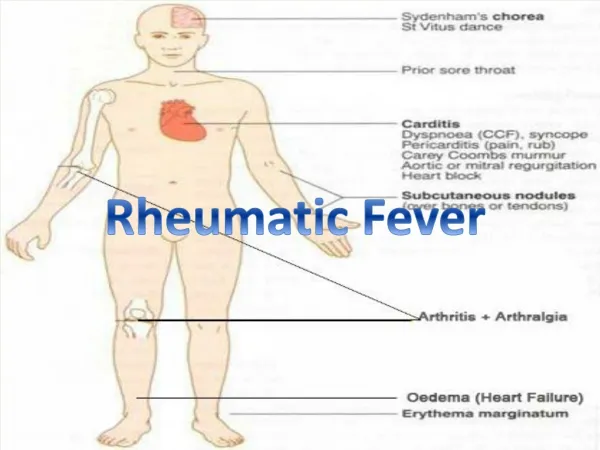
Carditis • Hallmark – Aschoff bodies • Found in all layers- endocardium, myocardium and pericardium
Aschoff body • Around 1-2mm • Lymphocytes, macrophages, Anitschkow cells and giant cells (Aschoff cells) • No myocytes • Perivascular • Goes through various stages Fibrinoid/Exudativestage (2-3 weeks) Granuloma/Proliferative stage(1-6months) Perivascularscarring • Anitschkowcells- caterpillar nucleus – giant macrophages • Aschoff cells – coalition of Anitschkow cells
Mc Callum’s patch McCallum’s patch: Gross finding of endocardial thickening in the posterior wall of LA due to inflammation as well as ‘jet’-trauma
CNS - CHOREA • Disseminated meningoencephalitis affecting basal ganglia, caudate nucleus, putamen, internal capsule and cerebellum • Obliterative endarteritis of cerebral and meningeal small vessels • Perivascular inflammation and petechial haemorrhage • Grossly normal brain tissue
ARTHRITIS • Endothelial inflammation of the synovia • Fibrinoidgranuloma, edema and diffuse inflammation. • Lasts for around 2-3 weeks • No permanent damage
SUBCUTANEOUS NODULE • Central zone of necrosis surrounded by surrounded by histiocytes and fibroblasts along with perivascular inflammation • Induration occurs principally due to perivascular oozing of plasma and cells into connective tissue. • Do not exhibit the pallisading pattern of RA
ERYTHEMA MARGINATUM • Dermal inflammation with minimal keratinocyte necrosis
Revised Jones criteria, 1965 • Supporting Evidence Of : • Preceeding Streptococcal Infection • H/O Recent Scarlet Fever; • Positive Throat C/S For Group A Streptococcus; • Increased ASO Titre
Jones criteria update, 1992 Supporting Evidence Of Preceeding Streptococcal Infection: Positive Throat C/S For Group A Streptococcus Rapid Antigen Test Elevated or Rising Streptococcal Antibody Titre
Concerns with Jones criteria • Did not cover – recurrence of RF indolent carditis chorea • Underdiagnosis in endemic area