Download
1 / 17
170 likes | 310 Vues
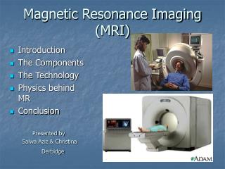
Practice Advisory on Anesthetic Care for Magnetic Resonance Imaging Anesthesiology 2009; 110:459 – 79 마취통증의학과 R4 최정현. -Introduction. Magnetic Rresonance Imaging (MRI) suite hazardous location presence of a strong static magnetic field

E N D
Practice Advisory on Anesthetic Care for Magnetic Resonance Imaging Anesthesiology 2009; 110:459–79 마취통증의학과 R4 최정현
-Introduction • Magnetic Rresonance Imaging (MRI) suite • hazardous location presence of • a strong static magnetic field • high-frequency electromagnetic(radiofrequency) waves • time-varied (pulsed) magnetic field • Secondary dangers • high-level acoustic noise • systemic and localized heating • accidental projectiles • significant challenges to anesthetic administration and monitoring capabilities • static and dynamic magnetic fields as well as radiofrequency energy emissions • Direct patient observation may be compromised by noise • darkened environment • obstructed line of sight • Distractions • requires broader responsibility for immediate patient care decisions
Methodology • Definition of Anesthetic Care for MRI and High-risk Imaging • Anesthetic care • moderate sedation • deep sedation • monitored anesthesia care • general anesthesia • ventilatory and critical care support • High-risk imaging refers • imaging in patients with medical or health-related risks • imaging with equipment-related risks • procedure-related risks • MRI-guided surgery • minimally invasive procedures : focused ultrasound, radiofrequency ablation • cardiac and airway imaging studies
Purpose of this advisory • (1) promote patient and staff safetyin the MRI environment • (2) prevent the occurrence of MRI-associated accidents • (3) promote optimal patient managementand reduce adverse patient outcomes associated with MRI • (4) identify potential equipment-related hazardsin the MRI environment • (5) identify limitations of physiologic monitoringcapabilities in the MRI environment • (6) identify potential health hazards(e.g., high decibel levels) of the MRI environment
-Education • All anesthesiologists should have general safety education • unique physical environment of the MRI scanner • specific features of individual scanners within their institution • emphasize safety for entering zones III and IV • hazards in this environment • effects on monitoring capabilities • should include information regarding • ferromagnetic items • stethoscopes, pens, wallets, watches, hair clips, name tags, pagers, cell phones, credit cards, batteries • implantable devices • spinal cord stimulators, implanted objects • should not be brought into zone III or IV
-Patient Screening • For every case, the anesthesiologist should communicate with • patient, referring physician, and radiologist • Whether the patient presents with a high-risk medical condition • neonatal status or prematurity • intensive or critical care status • impaired respiratory function • hemodynamic instability • vasoactive infusion requirements • comorbidities such as obesity and peripheral vascular disease • Requires equipment • physiologic or invasive monitors, intubation, oxygenation, or ventilation equipment • Has been screened for implanted devices • pacemakers, cardioverter–defibrillators, nerve stimulators • Has been screened for implanted ferromagnetic items • Surgical clips, prosthetic heart valves • Has been screened for the presence of imbedded foreign bodies • orbital iron filings, eyeliner tattoos
Patient Screening • anesthesiologist should communicate with the technologist to ensure that the patient has been screened for the presence of foreign bodies on the patient • (e.g., pierced jewelry, rings) before entering zone III • For patients with acute or severe renal insufficiency • should not administer gadolinium • because of the increased risk of nephrogenic systemic fibrosis • Cardiac pacemakers ,implantable cardioverter–defibrillators • generally contraindicated for MRI • When MRI is considered essential • should be developed in collaboration with the ordering physician, medical director or on-site radiologist, and other appropriate consultants (e.g., patient’s pacemaker specialist or cardiologist, diagnostic radiologist, device manufacturer).
Patient Screening • Certain implanted electronic devices • deep brain stimulators • Vagal nerve stimulators • phrenic nerve stimulators • wire-containing thermodilution catheters • cochlear implants • In consultation with • referring physician • radiologist responsible for the procedure • neurosurgeon • anesthesiologist • determined to be MRI safe/conditional before imaging of these patients
-Preparation • plan for providing optimal anesthetic care (1) requirements of the scan and personnel needs (2) positioning of equipment (3) special requirements or unique issues of patient or imaging study (4) positioning of the anesthesiologist and the patient (5) planning for emergencies • anesthesiologist should communicate with the radiology personnel • to determine the requirements for the scan • duration of the scan • position of the patient or area of the body in the scanner • positioning of receiver coils • need for periods of paused respiration • anesthesiologist should communicate with other anesthesia team members • regarding individual roles for anesthetic care • anesthesiologist should collaborate with the MR technologist and/or facility biomedical engineer • to determine optimal and safe location of movable equipment
Preparation Anesthesiologists should have (1) a clear line of sight of the patient and physiologic monitors (2) anesthetic delivery equipment located for optimal control of anesthetic depth and rapid intervention (3) access to hospital information systems integral to patient care • in the event of an emergency prepare a plan for rapidly summoning additional personnel • anesthesiologist should ensure (1) emergency equipment and drugs : immediately accessible (2) emergency communication (e.g.,phone or code button) : immediately available (3) evacuation plan : appropriate location outside the scan room (zone IV) for resuscitation • complete with physiologic monitors, oxygen, suction, and other appropriate resuscitation equipment
Patient Management during MRI < Monitoring > • ASA Standards for Basic Anesthetic Monitoring • Limitations of available monitoring equipment - electrocardiograms • superimposed voltages from blood flow in the high magnetic field • ST-segment interpretation may be unreliable, even with highly filtered monitors • When the anesthesia care provider is not in zone IV monitor should be available to view vital signs from zone III
Patient Management during MRI < Anesthetic care > • lighter levels of anesthesia may be appropriate during an MRI scan • lighter levels may result in airway complications • Laryngospasm • Coughing • other airway compromise • receiving deep sedation, ventilation cannot be directly observed during moderate sedation Monitoring of exhaled carbon dioxide should be considered • Monitoring oxygenation by pulse oximetry not a substitute for monitoring ventilatory function
Patient Management during MRI • Equipment (1) an integrated anesthesia machine, medical gases, and waste anesthesia gas disposal or gas scavenging • when inhalational anesthesia is administered (2) suction (3) adequate electrical outlets and lighting (4) storage areas for equipment and drugs • Equipment used in the MRI suite appropriate for the age and size of the patient • Although an anesthesia machine may not be required for the administration of total intravenous anesthesia must be equipment immediately available for the administration of positive pressure ventilation with oxygen
Patient Management during MRI < Airway management > • common airway problems • obstruction, secretions, laryngospasm, apnea and hypoventilation • risk for airway compromise more aggressive airway management • use of a endotracheal tube or laryngeal mask airway • may be less accessible when the patient is in the scanner • complex airway management (e.g., fiberoptic intubation) should be performed in a controlled environment outside of zone IV
Management of Emergencies • When a patient has a medical emergency (e.g., cardiopulmonary arrest) (1) Immediately remove the patient from zone IV while initiating cardiopulmonary resuscitation, if indicated (2) call for help (3) transport to safe area for resuscitation that is not in zone IV • close to zone IV as possible not to delay resuscitation efforts • contain resuscitation equipment • defibrillator • vital signs monitors • code cart : resuscitation drugs, airway equipment, oxygen, suction
Management of Emergencies • In the case of projectile emergencies • If possible, immediately remove the patient from zone IV and discontinue the scan • If the patient is injured proceed with medical emergency management • controlled quench may be necessary to remove the patient from the bore • When a quench occurs (1) immediately remove the patient from zone IV (2) immediately administer oxygen to the patient • Powerful static magnetic fields may persist after a quench therefore the usual precautions apply when entering zone IV
Postprocedure Care • Patients receiving sedation or anesthesia within the MRI suite access to postanesthetic care consistent • recovery rooms • dedicated intensive care • recovery areas within the MRI suite • intensive care and recovery areas • should include • vital sign monitors • oxygen • suction • resuscitation equipment • trained personnel • Patients should be given oral and written discharge instructions