Download

1 / 46
470 likes | 636 Vues
RADIATION PROTECTION. FORMAT:- RADIATION HAZARDS. Introduction Theories of radiation injury Factors affecting biologic tissues Radiation effects RADIATION PROTECTIONS RADIATION SOURCES RADIATION PROTECTION FOR THE OPERATOR FOR THE PATIENT FOR THE ENVIRONMENT

E N D
FORMAT:-RADIATION HAZARDS • Introduction • Theories of radiation injury • Factors affecting biologic tissues • Radiation effects RADIATION PROTECTIONS • RADIATION SOURCES • RADIATION PROTECTION FOR THE OPERATOR FOR THE PATIENT FOR THE ENVIRONMENT • RADIATION MONITORING DEVICES • RADIATION PROTECTION DEVICES • CONCLUSION • REFERENCES
INTRODUCTION: Spontaneous decay of radioactive materials produces radiation Radiation biology is defined as the study of effect of ionizing radiation on living system.
THEORIES OF RADIATION INJURY • Radiation act on living system through direct & indirect effects. when the energy of a photon or secondary electron ionize biologic macromolecule it is termed as direct effects. Alternatively , the photon may be absorbed by water in an organism , ionizing water molecules. The resulting ions form free radicals that in turn interact and produce change in biologic molecules. This series of event is termed as indirect effects.
SEQUENCE OF RADIATION INJURY Latent period : • The time that elapses between exposure to ionizing radiation and the appearance of observable clinical signs. • More the radiation received-shorter is the latent period. Period of injury: • Changes in cell function, breaking of chromosomes, cessation of mitotic activity or abnormal mitotic activity , cell death.
Recovery period: • Repair of radiation damage. • Lower dose of radiation more capacity to repair. • Damage due to high dose of radiation- not completely repaired.
RADIATION EFFECTS • Deterministic effects: it is an effect in which severity of the response is proportional to the dose. These have a dose threshold below which response is not seen. • Eg:Post radiotherapy mucositis
Stochastic effect: it is that effect for which the probability of the occurrence of a change, rather than its severity is dose dependent. These effect do not have a dose threshold. • Eg:Radiation induced cancer
Tissue and organ sensitivity • High Small lymphocytes Bone marrow Reproductive cells Intestinal mucosa • Intermediate Growing bone Salivary glands Lungs Kidney Liver • Low Muscle Optic lens
GENERAL EFFECTS OF RADIATION Skin: • Increased susceptibility to chapping. • Dryness and atrophy. • Progressive pigmentation. • Brittleness of finger nails. Hematopoietic injury: • Leukemia • Anemia • Lymphopenia
Eyes • Inflammation , fibrosis and decreased flexibility of the eyelid. • Damage to lacrimal glands-dryness. • Ulceration of the cornea. Ears • Radiation otitis media ( edema of mucosa and collection of fluid in middle ear) • Deafness due to rupture of ear drum.
SOURCES OF RADIATION EXPOSURE Can be divided into two: 1.Natural or background exposure. Natural exposure is radiation from natural sources 2.Artifical radiation. a) Medical 1) x-ray diagnosis 2) nuclear medicine b) Consumer products 1)occupational 2) nuclear fuel cycle fall out.
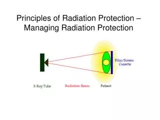
Sources of radiation in dental radiology department • Primary beam- radiation originating from focal sport. • Secondary radiation- radiation originating from the irradiated tissue patient. • Leakage or stray radiation- radiation from x-ray tube lead housing. • Scattered radiation- radiation coming from objects other than the patient as well as the wall and furniture's that primary beam strikes
PRINCIPLES FOR REDUCING DENTAL EXPOSURE 1.PRINCIPLE OF JUSTIFICATION To do more good than harm. Benefit to a patient from the diagnostic exposure exceeds the risk of harm. 2.PRINCIPLE OF OPTIMIZATION Reduce unnecessary exposure to patient. Principle of ALARA.
ALARA [As Low As Reasonably Achievable] concept:- 1)To avoid unnecessary exposure 2) Keep the dose as low as reasonably achievable 3) To ensure that these doses are justifiable
3)PRINCIPLE OF DOSE LIMITATION • Dose limits –no individuals are exposed to unacceptably high doses.
Three factors that control the dose or amount of radiation: • Time: • Reducing the time of an exposure reduces the effective dose proportionally. • Distance: • Increasing distance reduces dose due to the inversesquare law. • Shielding: • Adding shielding can also reduce radiation doses. The radiation getting through falls exponentially with the thickness of the shield.- Lead.
Radiation protection is the science of protecting people and the environment from the harmful effects of ionizing radiation. RADIATION PROTECTION CAN BE DIVIDED IN TO: • Protection for patient • Protection for operator • Protection for and from environment
Protection for patient • Patient protection techniques – Prior to exposure. During exposure After exposure
PRIOR TO EXPOSURE Proper prescribing of dental radiograph • Professional judgment of dentist is used to make decision about number, type and frequency. Proper Equipment • Dental x ray tube must be equipped with - Filters - Collimators - Position indicating devices
FILTRATION Low energy Rays are harmful and not useful in diagnostic radiography. INHERENT FILTRATION When the primary beam passes through x-ray tube. 0.5-1.0mm of aluminum. ADDED FILTRATION Placement of aluminum disks in the path of x-ray beam. Between collimator and tube head seal. Added as 0.5mm increments. Filter out longer wavelength .
TOTAL FILTRATION Sum of inherent and added filtration. At 70KVp -1.5mm Al. Above 70KVp -2.5mm Al.
COLLIMATION • Restricts the size and shape of x – ray beam and reduces patient exposure • Lead plate with hole in middle. It may be: • Rectangular • Round collimation: • Decreases the risk of radiation • Minimizes the scattered radiation • Sharper image with better contrast
POSITION INDICATING DEVICE • Extension of x ray tube head and used to directs the x-ray beam Three basic types • Conical • Rectangular • Round
PID’s 2 types based on length • Short (8inch) • Long(16inch) • X-rays are less divergent at a longer distance. Longer film spot to film distance results in 32 percent reduction in exposed tissue volume.
DURING EXPOSURE • Thyroid collars • Lead apron • Fast films • Intraoral image receptors • Film holding devices • Proper exposure factor selection • Good technique
THYROID COLLARS • Flexible lead shield placed around patient’s neck. • Protect thyroid from scatter radiation. • As a separate shield or as part of lead apron. • Recommended for all intraoral films. • Reduces exposure to gland by 92%.
LEAD APRON • Flexible shield that is placed over the patient chest and lap to protect the reproductive and blood forming tissues. • Recommended for all intraoral and extraoral films. • Reduces exposure – 98%.
FAST FILMS • Most effective method. • Reduces patient exposure. • F-speed film is the fastest film available • Fast films require less exposure bcoz film respond quickly due to fact that silver halide crystals in the emulsion are larger. INTENSIFYING SCREENSis device which transfer x rays into visible light, the visible light in turn expose the film. • 10-60 times more sensitive • Extra oral radiography • Emits green light with x rays
FILM HOLDING DEVICES • Reduces patient exposure. • Stabilize the film position in the mouth. • Provides external guide to indicate the film position. • Collimate the beam to a size of x ray film
AFTER EXPOSURE • Films must be handled and processed properly.Critical for the production of high-quality diagnostic radiographs. - Proper film handling • Necessary to produce diagnostic radiographs. • Limit patient exposure - Proper processing • Necessary for good radiographs. • Reduces retakes.; Reduce needless exposure. - Interpretation of image Images should be view under proper conditions with an illuminated viewer to obtain maximum available information.
PROTECTION FOR OPERATOR SOURCES OF EXPOSURE: • Primary x-ray beam. • Scattered radiation from filters, cones. • Scattered radiation from walls and furniture that the primary beam may strike. • Leakage radiation through the tube head housing.
Protection against primary beam • Effort must be made so that operator can leave the room or take suitable position behind the barriers. • Dental operatory should be designed and constructed to meet shielding requirements. • Lead apron is used.
Position Distance Rule: • Operator stands six feet away from source. • Should stand 90o to 135o with direction to central beam. • Film holding devices should be used. • Avoid holding the x-ray tube head
Protection against leakage radiation • Neither the tube nor the cone should be hand held. • Machine should be periodically checked for leakage. Protection against secondary or scattered radiation • High speed films • Open ended lead lined cone • Filtration • Collimator • Film badge/pocket dosimeter.
PROTECTION FOR THE ENVIRONMENT • Occupancy in the X ray room-Primary beam should be directed to patient • Assistance to patient- Holding of patient should be done by escort • Servicing of the unit- Certified & experienced persons • Quality assurance- Planned activity to ensure high quality images & minimum exposure
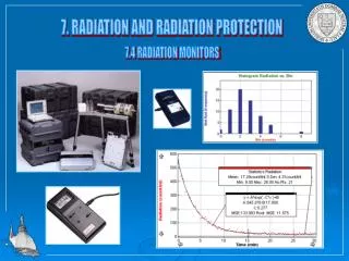
RADIATION MONITORING DEVICES • Measuring of X ray exposure Types • Electrical Ionization chamber Thimble chamber Proportional counter Geiger counter • Chemical Film Chemical dosimeter
Light Scintillation counter Gerenkov counter • Thermoluminescence Thermoluminescent Dosimeter • Heat Calorimeter
IONIZATION CHAMBER Consists of : • A pair of collecting plates each with an opposite charge. • Separated by a std volume of air. • Plates are connected by a electrometer. • The magnitude of the xray exposure is thus a function of the no:of ions produced and is measured by the magnitude of drop in potential between the collecting plates of the galvanometer
Personal monitoring devices Film badge • Plastic or metal housing – 12mm thick with a clip • Contains one or more film packets • Phenomenon: – x-rays blacken photographic film. Advantages • Measures any type and energy of radiation • Continuous assessment • Accumulated dose can be calculated • Permanent record • Simple and inexpensive
THERMOLUMINESCENCE DOSIMETER(TLD) • Actual dose received by operator or patient by radiography/radiotherapy exposures. • Easily placed on skin during exposure. • Principle-Thermo luminescence • Types of TL badges • Chest badge : used for estimation of the whole body dose, worn at the chest level. • Wrist badge: has a strap to be worn around wrist.
DOSE REPORTS :TLD badges are monitored every 3 months. • The dose reports are sent after processing the respective personal TL cards. • Doses are reported in units of mSv (millisievert). • Annual dose reports are sent after the end of the year and contain annual doses and cumulative life time doses of all radiation workers. • OVER EXPOSURE : • Dose equivalent recorded by the chest badge exceeding 100 mSv is treated as overexposure and the same is reported promptly to the institution and the individual concerned.
The institution should arrange to investigate the causes of overexposure and report the findings • a) The persons receiving more than 100 mSv will be subjected to hematological examination including differential blood counts and chromosome aberration test . • b)After receiving the investigation reports from the institution the overexposure cases are reviewed by BARC and advise on necessary follow-up will be intimated to the concerned institution.
BASIC PROTECTION REQUIREMENTS • Radiation area should be at one corner in the building such that at least two walls open to the environment. • Wall thickness in all directions equivalent to 2mm lead. • Doors should be protected by same thickness of lead. • All cracks & flanges around the door should be protected by additional strip of lead. • Windows should be at least 2m from the ground out side the x-ray room & at least 1.6m from the floor level inside the room. • Caution or warning signs should be displayed. • Waiting area should be outside x-ray room • Room adjacent office should be monitored.
REFERENCES • Oral radiology; principles and interpretation- White & Pharaoh. • Dental radiography- Haring & Jansen. • Text book of dental and maxillofacial radiology -Feny karjodkar. Principles of dental imaging-Langland & Langlais. • Inernet • Journal articles.