Download
1 / 72
750 likes | 983 Vues
6. Radiation Protection. Objectives. Define the key words. Adopt the ALARA concept. Use the selection criteria guidelines to explain the need for prescribed radiographs.

E N D
6 Radiation Protection
Objectives • Define the key words. • Adopt the ALARA concept. • Use the selection criteria guidelines to explain the need for prescribed radiographs. • Explain the roles communication, working knowledge of quality radiographs, and education play in preventing unnecessary radiation exposure.
Objectives • Explain the roles technique and exposure choices play in preventing unnecessary radiation exposure. • Explain the function of the filter. • State the filtration requirements for an intraoral dental x-ray unit that operates above and below 70 kVp.
Objectives • Compare inherent, added, and total filtration. • State the federally mandated diameter of the intraoral dental x-ray beam at the patient’s skin. • Explain the difference between round and rectangular collimation. • List the two functions of a collimator.
Objectives • Explain how PID shape and length contribute to reducing patient radiation exposure. • Identify film speeds currently available for dental radiography use. • Explain the role image receptor holders play in reducing patient radiation exposure.
Objectives • Advocate the use of the lead/lead equivalent thyroid collar and apron. • Explain the role darkroom protocol and film handling play in reducing patient radiation exposure. • Summarize the radiation protection methods for the patient.
Objectives • Explain the roles time, shielding, and distance play in protecting the radiographer from unnecessary radiation exposure. • Utilize distance and location to take a position the appropriate distance and angle from the x-ray source at the patient’s head during an exposure.
Objectives • Describe monitoring devices used to detect radiation. • Summarize the radiation protection methods for the radiographer.
Objectives • List the organizations responsible for recommending and setting exposure limits. • State the maximum permissible dose (MPD) for radiation workers and for the general public.
Key Words • Added filtration • ALARA (as low as reasonably achievable) • Aluminum equivalent • Area monitoring • Collimation
Key Words • DIS (direct ion storage) monitor • Dosimeter • Exposure factors • Film badge • Film/image receptor holder • Filter
Key Words Filtration Half-value layer Inherent filtration Lead apron Lead equivalent MPD (maximum permissible dose) Monitoring
Key Words OSL (optically stimulated luminescence) monitor Personnel monitoring Personnel monitoring device
Key Words • PID (position indicating device)/BID (beam indicating device) • Primary beam • Protective barrier • Radiation leakage • Radiation workers
Key Words • Retake radiographs • Scatter (secondary) radiation • Selection criteria • Structural shielding • Thermoluminescent dosimeter (TLD) • Thyroid collar • Total filtratio
Introduction • Although it is the consensus of radiobiologists that the dose received from a dental x-ray exposure is not likely to be harmful, even the experts do not know what risk a small dose carries.
Introduction • The patient has agreed to be subjected to the risks of radiation exposure because he/she believes that the oral health care practitioner will follow safety protocols that protect the patient from excess exposure.
Introduction • In this chapter, we discuss radiation safety protocols, including selection criteria used in prescribing dental radiographs and methods to minimize x-ray exposure to both the dental patient and the radiographer.
ALARA “As Low As Reasonably Achievable”
Protection Measures for the Patient • Professional judgment • Technical ability of the operator • Technique standards • Equipment standards • Optimum film processing
Protection Measuresfor the Patient • Professional Judgment • Use evidence-based selection criteria when determining which patients need radiographs. • Guidelines adopted by the American Dental Association that assist in deciding when, what type, and how many radiographs should be taken are shown in Table 6-1.
Protection Measuresfor the Patient • Technical Ability of the Operator • Communication — clear instructions; patient cooperation • Working knowledge of quality radiographs — understand what a quality dental radiograph should image • Education — continuing education is important
Protection Measuresfor the Patient • Technique Standards • Intraoral technique choice — first choice; The paralleling technique yields more accurate and precisely sized radiographic images • Exposure factors — select the appropriate exposure factors — kilovoltage(kVp), milliamperage (mA), and time.
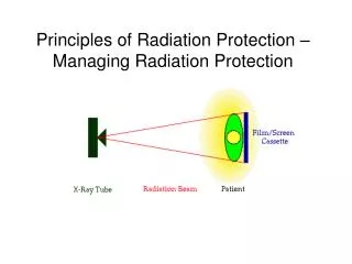
Protection Measuresfor the Patient • Equipment Standards: The Federal Performance Standard for Diagnostic X-ray Equipment, effective August 1974. • Provisions require that all x-ray equipment manufactured after this date meet certain radiation safety requirements including filtration, collimation, and PID
Figure 6-1 Collimator and filter. The collimator is a lead washer that restricts the size of the x-ray beam. The filter is an aluminum disc that filters (removes) the long wavelength x-rays.
Figure 6-2 Effect of filtration on skin exposure. Aluminum filters selectively absorb the long wavelength x-rays.
Figure 6-3 Effect of collimation on primary beam. Lead collimators control the shape and size of the primary beam. The beam is limited to the approximate size of the image receptor.
Figure 6-4 External collimator attaches to the PID to reduce the area of radiation exposure.
Figure 6-5 The collimator restricts the size of the primary beam to 2.75 in. (7 cm) at the end of the PID.
Figure 6-6 Although circular collimation provides a large enough area of exposure to adequately cover a size #2 image receptor, the patient also receives excess radiation not needed for the exposure of this receptor.
Figure 6-7 Rectangular PIDs restrict the x-ray beam to the approximate size of a #2 intraoral image receptor. Rectangular PIDs are available in 8, 12, and 16 inches (20.5, 30, and 41 cm). (Courtesy of Margraf Dental Manufacturing Inc.)
Figure 6-8 Plastic closed-ended, pointed “cones” are no longer used.
Figure 6-9 Round PIDs are available in 16, 12, and 8 inches (41, 30, and 20.5 cm).
Figure 6-10 Target-surface distance. The longer the target-surface distance, the more parallel the x-rays and the less tissue exposed. Note that the beam size at the patient’s skin entrance is 2.75 in. (7 cm) for both target-surfacedistances. It is the exit beam size that increases to expose a larger area when using the shorter target-surface distance.
Figure 6-11 Many image receptor holding devices are available to fit most situations. The use of a holder prevents asking patients to put their fingers in the path of the primary beam.
Figure 6-12 Lead aprons and thyroid collars are available in a wide range of sizes. Aprons are available with an attached thyroid collar, or the thyroid collar may be a separate part.
Figure 6-13 Patient protected with lead apron with thyroid collar in place.
Protection Measuresfor the Patient • Optimum Film Processing • Processing errors increase patient radiation exposure by resulting in retake radiographs. • Patient protection techniques should be used at all times to keep radiation exposures as low as possible
Protection Methods for the Radiographer • All measures taken to protect the patient from radiation also benefit the radiographer. • Radiation protection methods include time, shielding, and distance. • The radiographer should spend a minimal amount of time, protected by shielding, at the greatest distance from the source of radiation to avoid unnecessary exposure.
Figure 6-14 Distance is an effective means of reducing exposure from scatter radiation.
Figure 6-15 When structural shielding is not available, the radiographer should stand in a position at least 6 ft(1.83 m) from the head of the patient at an angle of 45º to the exiting primary beam.
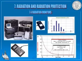
Radiation Monitoring • Area monitoring • Personnel monitoring: Types • Film badge • Thermoluminescent dosimeter (TLD) • OSL (optimally stimulated luminescence) monitor • DIS (direct ion stimulation) monitor
Figure 6-16 OSL radiation monitor worn bythe radiographer to monitor radiation exposure. (Courtesy of Landauer, Inc.)
Figure 6-17 DIS radiation monitor. Sized and shaped similar to a thumb drive, this device has a clip to allow the radiographer to wear the monitor while working with ionizing radiation. The device uses a USB connector to plug into a computer with Internet access. When logged on to the manufacturer’s Web site, real-time radiation exposure readings may be downloaded from the device. (Courtesy of Quantum Products.)