Download
1 / 38
380 likes | 630 Vues
Joints. Articulating your body. Joints (Articulations). Weakest parts of the skeleton Articulation – site where two or more bones meet Functions of joints Give the skeleton mobility Hold the skeleton together. Classification of Joints: Structural. Structural classification

E N D
Joints Articulating your body
Joints (Articulations) • Weakest parts of the skeleton • Articulation – site where two or more bones meet • Functions of joints • Give the skeleton mobility • Hold the skeleton together
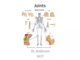
Classification of Joints: Structural • Structural classification • focuses on the material between bones • Whether or not a joint cavity is present • The three structural classifications are: • Fibrous • Cartilaginous • Synovial
Classification of Joints: Functional • Functional classification is based on the amount of movement allowed by the joint • The three functional classes of joints are: • Synarthroses – immovable • Amphiarthroses – slightly movable • Diarthroses – freely movable
Fibrous Structural Joints • The bones are joined by fibrous tissues • There is no joint cavity • Most are immovable • There are three types • Sutures • Syndesmoses • gomphoses
Sutures Figure 8.1a
Sutures • Occur between the bones of the skull • Comprised of interlocking junctions completely filled with connective tissue fibers • Bind bones tightly together, but allow for growth during youth • In middle age, skull bones fuse and are called synostoses
Syndesmoses Figure 8.1b
Syndesmoses • Bones are connected by a fibrous tissue ligament • Movement varies from immovable to slightly variable
Gomphoses • The peg-in-socket fibrous joint between a tooth and its alveolar socket • The fibrous connection is the periodontal ligament
Cartilaginous Joints • Articulating bones are united by cartilage • Lack a joint cavity • Two types – synchondroses and symphyses
Synchondroses Figure 8.2a, b
Synchondroses • A bar or plate of hyaline cartilage unites the bones • All synchondroses are synarthrotic
Symphyses Figure 8.2c
Symphyses • Hyaline cartilage covers the articulating surface of the bone and is fused to an intervening pad of fibrocartilage • Amphiarthrotic joints designed for strength and flexibility
Synovial Joints • Those joints in which the articulating bones are separated by a fluid-containing joint cavity • All are freely movable diarthroses • Examples – all limb joints, and most joints of the body
General Structure • Synovial joints all have the following • Articular cartilage • Joint (synovial) cavity • Articular capsule • Synovial fluid • Reinforcing ligaments
General Structure Figure 8.3a, b
Friction-Reducing Structures • Bursae – flattened, fibrous sacs lined with synovial membranes and containing synovial fluid • Common where ligaments, muscles, skin, tendons, or bones rub together • Tendon sheath – elongated bursa that wraps completely around a tendon
Friction-Reducing Structures Figure 8.4
Range of Motion • Nonaxial – slipping movements only • Uniaxial – movement in one plane • Biaxial – movement in two planes • Multiaxial – movement in or around all three planes
Stability • Determined by: • Articular surfaces • shape determines what movements are possible • Ligaments • unite bones and prevent excessive or undesirable motion • Muscle tone is accomplished by: • Muscle tendons across joints acting as stabilizing factors • Tendons that are kept tight at all times by muscle tone
Range of Motion • Nonaxial • slipping movements only • Uniaxial • movement in one plane • Biaxial • movement in two planes • Multiaxial • movement in or around all three planes
Gliding Movements • One flat bone surface glides or slips over another similar surface • Examples – intercarpal and intertarsal joints, and between the flat articular processes of the vertebrae
Gliding Movement Figure 8.5a
Angular Movement • Flexion • bending movement that decreases the angle of the joint • Extension • reverse of flexion; joint angle is increased
Angular Movement Figure 8.5b
Angular Movement Figure 8.5c, d
Angular Movement • Dorsiflexion and plantar flexion • up and down movement of the foot • Abduction • movement away from the midline • Adduction • movement toward the midline • Circumduction • movement describes a cone in space
Angular Movement Figure 8.5e, f
Rotation • The turning of a bone around its own long axis • Examples • Between first two vertebrae • Hip and shoulder joints Figure 8.5g
Special Movements • Supination and pronation • Inversion and eversion • Protraction and retraction • Elevation and depression • Opposition
Special Movements Figure 8.6a
Special Movements Figure 8.6b
Special Movements Figure 8.6c
Special Movements Figure 8.6d
Special Movements Figure 8.6e