Download
1 / 8
80 likes | 89 Vues
BASIC HYPER SPECTRAL IMAGING . Fred Sigernes 1,2,3,4. 1 The University Centre in Svalbard (UNIS), N-9171 Longyearbyen, Norway 2 The Birkeland Centre for Space Science (BCSS) 3 The Kjell Henriksen Observatory (KHO) 4 Centre for Autonomous Marine Operations and Systems (AMOS) NTNU.

E N D
BASIC HYPER SPECTRAL IMAGING Fred Sigernes 1,2,3,4 1 The University Centre in Svalbard (UNIS), N-9171 Longyearbyen, Norway 2 The Birkeland Centre for Space Science (BCSS) 3 The Kjell Henriksen Observatory (KHO) 4 Centre for Autonomous Marine Operations and Systems (AMOS) NTNU Lectures: TTK20 Hyperspectral remote sensing, Module 1, AMOS – NTNU, 11-13 September, 2018.
Lecture plan: TTK20 Hyperspectral remote sensing Module 1 Instructor: Adjunct Professor Fred Sigernes Lectures: TTK20 Hyperspectral remote sensing, Module 1, AMOS – NTNU, 11-13 September, 2018.
3.1 Spectrometer optical diagram An optical diagram is a standard way to trace rays through a spectrometer in an unrolled linear fashion. It visualizes a center cross section of the instrument perpendicular to the slits, parallel to the axis of refraction.
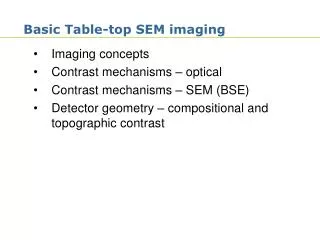
3.2 Front optics Single thin Lens equation Magnification Numerical aperture F/value or f/#
3.3 f/value of a spectrometer The f/value of s spectrometers depends on whether you watch the grating from the exit slit or the entrance slit. Define D as equivalent diameter as seen from slits. The f/values are then
Slit width magnification is different and is given as This will be proven later! 3.4 Magnification of the slits Slit height magnification from single lens consideration gives The grating acts as lens along the entrance slit of height h. Slit height magnification is then simply the result of a lens with object distance f2 and image distance f3.
3.5 Bandpass and resolution Bandpass is the measure of an instrument ability to separate adjacent spectral lines. It is defined as the recorded Full Width at Half Maximum (FWHM) of a monochromatic spectral line If F is the recorded spectrum, B the source spectrum and P the instrumental line profile then - related to width of the slits, natural line width, resolution, alignment, diffraction effects, aberrations or quality optics etc.
Assume resolution depends on width of slits and that the instrument is perfectly aligned.