Download

1 / 44
440 likes | 748 Vues
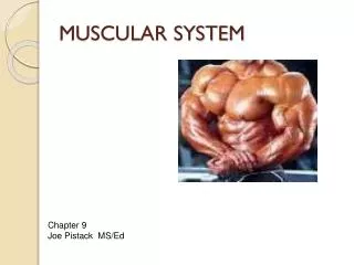
Muscular System: Chapter 8. Chapter 8. Functions of Muscles . 1) Movement Move the skeleton Move food and body fluids Create heartbeat 2) Heat Production Used to regulate body temperature. Types of Muscle Tissue. 1) Skeletal Striated, Voluntary Multiple nuclei/cell

E N D
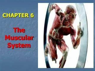
Muscular System:Chapter 8 Chapter 8
Functions of Muscles • 1) Movement • Move the skeleton • Move food and body fluids • Create heartbeat • 2) Heat Production • Used to regulate body temperature
Types of Muscle Tissue • 1) Skeletal • Striated, Voluntary • Multiple nuclei/cell • Ex: Quadriceps, triceps • 2) Cardiac • Striated, Involuntary • 1 nucleus/cell • Ex: heart • 3) Smooth • Unstriated, Involuntary • 1 nucleus/cell • Ex: stomach wall, espophagus
Structure of a Muscle • Fascia • Outer layer of fibrous connective tissue • Continuous with tendon and/or bone • Epimysium • Layer under the fascia • Perimysium • Layer under epimysium • Wraps around bundles called fascicles
Structure of a Muscle (cont) • Endomysium • Layer under perimysium • Wraps around muscle fiber • Sarcolemma • Layer under endomysium • Cell membrane of a muscle cell (fiber) • Surrounds bundles of myofibrils
Structure of a Muscle Fiber • Sarcolemma- cell membrane • Sarcoplasm- cytoplasm • Sarcoplasmic reticulum- endoplasmic reticulum • Multiple nuclei/cell • Many mitochondria • Transverse tubules- membrane-bound canals through the fiber; • surrounded by cisternae of sarcoplasmic reticulum • Filled with bundles of myofibrils
Structure of the Myofibril • Composed of myosin (thick) filaments and actin (thin) filaments • Filaments overlap creating striations • Z-line- attachment for actin filaments • M-line- attachment for myosin filaments • I-band- zone containing only actin filaments • A-band- zone containing myosin filaments • H-zone- zone containing only myosin filaments • Sarcomere- unit stretching from one Z-line to the next
Neuromuscular Junction • Motor neuron - nerve that connects to muscle fiber • Neuromuscular junction - connection between nerve and muscle fiber • Motor end plate - specialized area of the sarcolemma modified to connect with the nerve • Neurotransmitters - messengers that are stored in synaptic vesicles in the neuron and released across synaptic cleft
Motor Units • A fiber usually has 1 neuromuscular junction • A motor neuron can be connected to many fibers • Motor unit - a motor neuron and all of its connected fibers • Fibers will contract as a unit
Quick Review • If your muscle cells were not producing enough ATP, which part of the cell is dysfunctional? • A) Sarcoplasmic reticulum • B) Sarcolemma • C) Mitochondria • D) Nucleus
Quick Review • If you were diagnosed with a disease that affected your ability for your muscles to communicate (connect) to your nervous tissue, which part of your muscle would this affect? • A) Motor unit • B) Motor neuron • C) Neuromuscular junction • D) All of the above
Sliding Filament Model • Structure animation • Muscle shortens as filaments slide past each other • This means that the I-band will get smaller during a contraction
Timeline of a Contraction • Step 1- Release of Acetylcholine • Acetylcholine is a neurotransmitter made and stored in the neuron • Release with nerve impulse into synaptic cleft • Crosses cleft and binds with receptors on motor plate • Step 2- Muscle Impulse • Binding of acetylcholine at motor plate stimulates muscle impulse • Impulse spreads across sarcolemma and down into T-tubules
Timeline of a Contraction (cont) • Step 3- Movement of Calcium • Cisternae of sarcoplasmic reticulum become more permeable to Ca+ ions • Ca moves out of reticulum into sarcoplasm • Step 4- Exposing Binding Sites of Actin • High Ca+ in sarcoplasm cause a change in the actin filaments • Troponin and tropomyosin • Thin filaments attached to actin; act together to expose the binding site
Timeline of a Contraction (cont) • Step 5- Contraction • Readied myosin heads attach to exposed actin binding sites and pull • A new ATP must bind with the myosin ATPase before myosin will release binding site • Readied myosin head then binds with a new actin binding site • I-band gets smaller • This will continue as long as acetylcholine is present
Timeline of a Contraction (cont) • Step 6- Relaxation • Two steps lead to relaxation: • 1) Acetylcholinesterase breaks down acetylcholine • 2) Once acetylcholine is low, Ca+ is actively pumped back into sarcoplasmic reticulum • Low Ca+ levels in sarcoplasmstop linkage of actin and myosin and muscle fiber relaxes to it normal length Muscle Contraction Animation: Myofibril Muscle Contraction Animation: Sarcomere
Quick Review • Which protein filaments are involved in muscle contraction? • A. Actin • B. Myosin • C. ATPase • D. More than one answer is correct
Quick Review • Which muscle fiber structures are involved in contraction? • A. I-band • B. Sarcomere • C. Active site • D. More than one answer is correct
Quick Review • Acetycholine is a neurotransmitter whose amount will increase during contraction (to a point); the amount then decreases to stimulate relaxation. • True • False
Energy Sources for Contraction • 1st source- available ATP’s (very small amount) • 2nd source- Creatine phosphate breaks down to produce more ATP • 3rd source- Cellular respiration to create new ATP’s • Extra oxygen stored in myoglobin in muscles • 4th source- Anarobic respiration • Creates a build-up of lactic acid
Oxygen Debt • Lactic Acid is moved to the liver to be converted back to glucose • Oxygen debt • Amount of oxygen needed for liver to convert the lactic acid • How much is needed by the muscle to reset the other sources • Debt may take hours to repay after strenuous activity
Muscle Fatigue • Occurs because: • Blood supply interrupted • Acetylcholine used up • Build-up of lactic acid which lower pH of muscle which lowers muscles response to stimulation
Muscle Fiber Responses • Threshold stimulus - intensity of stimulation needed to make a contraction occur • All-or-none response - muscle fiber responds fully or not at all
Recording Muscle Fiber Contractions • Recording is a myogram • Latent period- period of time between stimulus and response • Period of Contraction • Period of Relaxation • Making a muscle fiber go through a single contraction is called a twitch
Quick Review • Muscles could take hours to recover from oxygen debt. • True • False
Quick Review • Which of the following is a reason why a muscle could become fatigued? • A. Blood supply increases • B. Acetylcholine is present • C. Build-up of lactic acid which lowers pH of muscle • D. None of the above
Summation and Tetany • Summation - strength of muscle fiber response increases if another stimulus is applied before relaxation is finished • Tetany - a sustained maximum muscle fiber response produced by a high frequency of stimuli that don’t allow the muscle to relax
Recruitment • Muscles do NOT have all-or-none contractions • Muscles are made of many motor units • Respond to a variety of stimulus strengths • Muscle used for strength normally have more bigger motor units • Muscles used for fine movements have more smaller motor units
Muscle Tone • A few motor units go through sustained contractions • Help keep posture and support
Skeletal Muscle Action • Origin - muscle attachment on bone that is immobile during movement • Insertion- muscle attachment on bone that will move • For any body movement: • Prime mover (agonist)- major muscle creating movement • Synergist- help with movement • Antagonist – create movement in the opposite direction
Smooth Muscle • Contains myosin and actin filaments but more randomly arranged (no striations) • Multiunit- stimulus is through nerves or hormones (iris or walls of blood vessels) • Visceral- cells can stimulate each other (walls of intestine, uterus, urinary tract) • Peristalsis- wave-like contraction
Cardiac Muscle • Cells form interconnecting network • Cells are connected at intercalated disks • Impulses can rapidly transmit from cell to cell • Network response is all-or-none
Don’t forget: You can copy-paste this slide into other presentations, and move or resize the poll.
Animation and Quiz! • Animation with Quiz: http://bcs.whfreeman.com/thelifewire/content/chp47/4702001.html