Download
1 / 188
1.96k likes | 2.35k Vues
CHAPTER 6 The Muscular System. SECTION 1: OVERVIEW OF MUSCLE AND THE MUSCULAR SYSTEM. Primary Functions of the Muscular System. 1.) Producing movement 2.) Maintaining posture 3.) Stabilizing joints and providing structure and support for organs and tissues 4.) Generating heat.

E N D
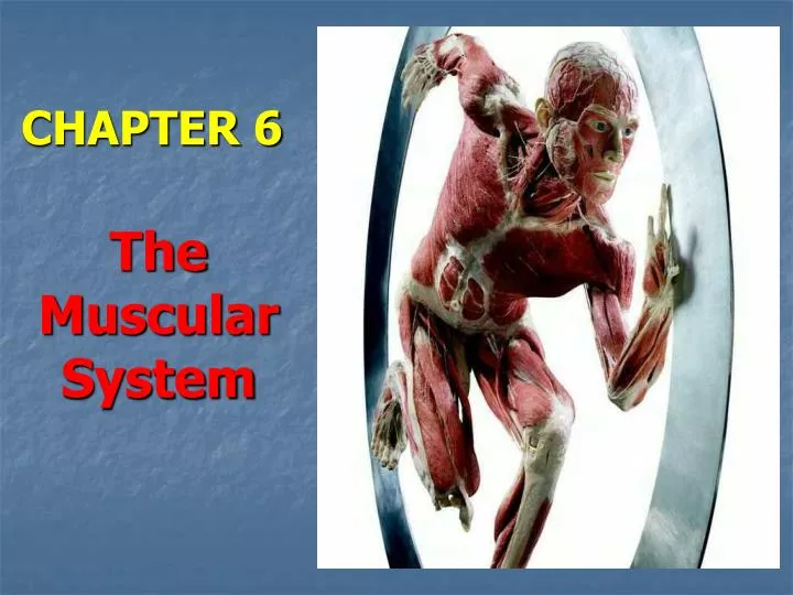
CHAPTER 6 The Muscular System
Primary Functions of the Muscular System • 1.) Producing movement • 2.) Maintaining posture • 3.) Stabilizing joints and providing structure and support for organs and tissues • 4.) Generating heat
Overview of Muscle Tissues • The essential function of muscle is contraction or shortening – a unique characteristic that sets it apart from other body tissue. • All muscles cells are elongated and called muscle fibers. • The ability of muscles to shorten, or contract, depends on two types of myofilaments --filaments composing the myofibrils, which are the contractile organelles in the cytoplasm of muscle cells. There are two types of myofilaments: actin and myosin. • Myo or mys means “muscle,” and sarco means “flesh”
Muscle Tissue: 3 Types • Muscle tissue enables the movement of body structures. • There are three types of muscle tissue: • smooth muscle tissue, • cardiac muscle tissue, and • skeletal muscle tissue.
Skeletal muscle is an example of voluntary muscle, and appears banded or striated. Skeletal muscle is sometimes called striated muscle, and tires after short periods of activity. • Smooth muscles are involuntary muscles that have no striations. Smooth muscles propel substances along a definite tract, or pathway. • Cardiac muscle is involuntary muscle whose cells are striated. Cardiac muscle is different from skeletal muscle in that it does not get tired.
cardiac muscle tissue intercalated discs
SKELETAL MUSCLE: BRIEF SUMMARY • voluntary muscle • banded or striated (sometimes called striated muscle) • cigar-shaped (cylindrical), long, multinucleate cells, and the largest of the muscle fiber types • found in skeletal muscles that attach to the body’s skeleton • functions to contract and relax to move skeletal bones • tires easily and must rest after short periods of activity
SMOOTH MUSCLE: BRIEF SUMMARY • involuntary muscle • no striations • spindle-shaped and have a single nucleus (uninucleate cells), arranged in sheets or layers • found mainly in the walls of hollow visceral organs – stomach, urinary bladder, interior walls of blood vessels, and respiratory passages • propels substances along a definite tract, or pathway • Smooth muscle contraction is slow and sustained.
CARDIAC MUSCLE: BRIEF SUMMARY • involuntary muscle found only in the heart • striated • cardiac fibers cushioned by soft connective tissue arranged in spiral or figure 8-shaped bundles • branching chains of uninucleate cellsjoined by junctions called intercalated discs • contracts at a fairly steady rate set by the heart’s pacemaker, but can be stimulated by the nervous system to shift to “high gear” for short periods • does not get tired
cardiac muscle tissue intercalated discs
Muscle Types: Skeletal Muscle • Skeletal muscle is striated, and associated with voluntary movement. • It also provides structure and support for organs and tissues.
Anatomy of Skeletal Muscles • The endomysium isa delicate, thin connective tissue sheath that surrounds each muscle cell. • The endomysium surrounds the individual skeletal muscle cells and loosely interconnects adjacent muscle fibers. • The endomysium is flexible and contains capillary networks and nerve fibers.
Figure 6.1 page 164 epimysium perimysium muscle fascicle tendon endomysium skeletal muscle skeletal muscle fiber (cell)
Anatomy of Skeletal Muscles • The perimysium is made of connective tissue fibers and divides the skeletal muscle into a series of compartments. • The perimysium contains blood vessels and nerves that maintain blood flow and innervate the muscle fibers within the fascicle. • Each compartment contains a bundle of muscle fibers bound together by connective tissue that is known as a fascicle. Many fascicles are bound together by an even tougher “overcoat” of connective tissue called an epimysium, which covers the entire muscle.
Figure 6.1 page 164 epimysium perimysium muscle fascicle tendon endomysium skeletal muscle skeletal muscle fiber (cell)
Figure 6.1 page 164 epimysium perimysium 3. 5. muscle fascicle tendon 4. 7. 6. endomysium 2. skeletal muscle skeletal muscle fiber (cell) 1.
The epimysium separates the muscle from surrounding tissues and organs. • The epimysium is connected to the deep fascia, a dense connective tissue layer. • The epimysia blend into the strong, cordlike tendons, or into fibrous membranous sheets called aponeuroses which attach muscles indirectly to bones, cartilages, or connective tissue coverings of each other.
Tendons serve to anchor muscles to bones, but also provide durability, and conserve space. • Tendons are mostly tough collagenic fibers, so they can cross rough bony projections without tearing. • Because of their relatively small size, more tendons than fleshy muscles can pass over a joint. • Muscles vary considerably in the way their fibers a arranged. Many are spindle-shaped, but in others the fibers are arranged in a fan shape or in a circle.
Looking back at the anatomy of a muscle cell, and placing the structures in order from largest to smallest gives this arrangement: • a fascicle – a bundle of muscle fibers made up of individual • muscle fibers that are composed of • myofibrils – organelles found in the cytoplasm of muscle cells that are made up of tiny units called • sarcomeres -- composed of chains of tiny contractile units called • myofilaments – threadlike protein filaments of actin or myosin
Muscle Types: Smooth Muscle • Smooth muscle is non-striated, and acts in a number of involuntary processes in the body. • Smooth muscles is found mainly in the walls of hollow visceral organs such as the stomach, urinary bladder, and respiratory passages. • Smooth muscles helps to propel substances along a definite tract, or pathway, within the body.
Muscle Types: Smooth Muscle • Smooth muscle cells are spindle-shaped and have a single nucleus (uninucleate). • Smooth muscle cells are arranged in sheets or layers. Most often, there are two layers, one running circularly and the other longitudinally. • As the two layers alternately contract, and relax, they change the size and shape of the organ. • Smooth muscle contraction is slow and sustained.
Peristalsisis the wavelike contractions seen in tube-like organs that propels substances along a tract.
Muscle Types: Smooth Muscle Some of the involuntary processes of smooth muscle include: • allows the expansion and contraction of arteries and veins • lines the bladder and reproductive tracts • lines the entire gastrointestinal tract
Did you know... • Tiny smooth muscle fibers in the skin called arrector pili are responsible for “goose bumps.”
Muscle Types: Cardiac Muscle • Cardiac muscle(heart muscle) is striated but functions involuntarily. • It is solely responsible for propelling blood throughout the body. • Cardiac muscle fibers are branching cells joined by special junctions called intercalated discs. • The heart’s pacemaker is tissue that sets and maintains the heart’s steady rate. • Unlike other muscle types, cardiac muscle does not tire.
Cardiac fibers are cushioned by small amounts of soft connective tissue and arranged in spiral or figure 8-shaped bundles. • Cardiac muscle fibers are branching cells joined by special anchoring structures called intercalated discs that contain gap junctions. • These two structural features and the spiral arrangement of the muscle bundles in the heart allow heart activity to be closely coordinated.
is involuntary muscle smooth muscle is involuntary muscle is involuntary muscle is voluntary muscle is voluntary muscle cardiac muscle is voluntary muscle has striations has striations has striations skeletal muscle does not have striations does not have striations does not have striations
Microscopic Anatomy of Skeletal Muscle • The sarcolemma is the cell membrane of a muscle cell that is found in skeletal, cardiac, and smooth muscle. • A sarcolemma consists of a true cell membrane, called the plasma membrane, and an outer coat made up of a thin layer of polysaccharide material that contains numerous thin collagen fibrils. • At each end of the muscle fiber, this surface layer of the sarcolemma fuses with a tendon fiber, and the tendon fibers in turn collect into bundles to form the muscle tendons that then insert into bones. • The membrane is configured to receive and conductstimuli.
Microscopic Anatomy of Skeletal Muscle • Myofibrils are small cylindrical, contractile organelles found in the cytoplasm of muscle cells. • Alternating light (I)bands and dark (A) bands along the length of the perfectly aligned myofibrils give the muscle cell as a whole its striped appearance. • The light I bandhas a midline interruption, a darker area called the Z disc. • The dark A bandhas a lighter central area called the H zone.
A myofibril consists of approximately 10,000 sarcomeres end to end. • A sarcomere is the functional component within the myofibril that is composed of chains of tiny contractile units called myofilaments. • Sarcomeres are aligned end-to-end along the length of the myofibrils. A sarcomere lies in the area between the two Z lines. • Within the sarcomere, there are two types of threadlike protein myofilaments within each of the sarcomeres: actinand myosin. • The protein filaments actin and myosin allow the cell to expand and contract in a three-step process.
Myosin Filaments • Largerthick filaments, or myosin filaments, are the thick filaments made mostly of bundled molecules of the protein myosin. • Myosin contains ATPase enzymes, which split ATP to generate the power for muscle contraction. • The midpoints of the thick filaments are smooth, but their ends are studded with small projections called myosin heads, which aresometimes called cross bridgesbecause they link the thick and thin filaments together during contraction.