Download
1 / 54
600 likes | 813 Vues
Urinary System. Ch 25. Function. Remove nitrogenous wastes Maintain electrolyte, acid-base, and fluid balance of blood Homeostatic organ Acts as blood filter Release hormones: calcitriol & erythropoietin. Kidneys as Filters. Diuretic- lose water; coffee, alcohol

E N D
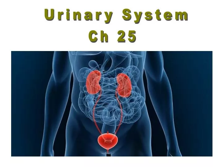
Urinary System Ch 25
Function • Remove nitrogenous wastes • Maintain electrolyte, acid-base, and fluid balance of blood • Homeostatic organ • Acts as blood filter • Release hormones: calcitriol & erythropoietin
Kidneys as Filters • Diuretic- lose water; coffee, alcohol • Antidiuretic- retain water; ADH • Aldosterone- sodium & water reabsorption, and K+ excretion • GFR= 180 liters (50 gal) of blood/day • 178-179 liters are reabsorbed back into blood • Excrete a protein free filtrate
Renal Artery • Segmental Arteries • Interlobar Arteries • Arcuate Arteries • Cortical Radiate Arteries • Afferent Arterioles • Glomerular Capillaries • Efferent Arterioles • Peritubular Capillaries • Cortical Radiate Vein • Arcuate Veins • Interlobar Veins • Renal Veins
Nephron • consist of a long tubule & a ball of capillaries called a glomerulus • the end of the tubule that surrounds the glomerulus is called Bowman’s capsule • the remaining parts of the tubule are called the: • proximal tubule • loop of Henle • distal tubule • the tubule empties into a collecting duct that leads to the renal pelvis • the renal pelvis opens to the ureter
Nephron Each kidney contains over 1 million nephrons and thousands of collecting ducts
Nephron’s functions: • glomerular filtration • tubular reabsorption • tubular secretion
Composition of Glomerular Filtrate • Water • Small Soluble Organic Molecules • Mineral Ions
Boman’s Capsule • filtration occurs as blood pressure in the capillaries of the glomerulus forces filtrate into Bowman’s capsule • the process is passive (diffusion) • the filtrate includes: • water, salts, bicarbonate (HCO3–), H+, urea, glucose, amino acids
Proximal Convoluted Tubule Reabsorbs: water, glucose, amino acids, and sodium. • 65% of Na+ is reabsorbed • 65% of H2O is reabsorbed • 90% of filtered bicarbonate (HCO3-) • 50% of Cl- and K+
Loop of Henle Creates a gradient of increasing sodium ion concentration towards the end of the loop within the interstitial fluid of the renal pyramid. • 25% Na+ is reabsorbed in the loop • 15% water is reabsorbed in the loop • 40% K is reabsorbed in the loop
Loop of Henle:Descending Limb • is impermeable to salts but permeable to water • as filtrate moves through the descending limb, water steadily moves out making the filtrate more & more concentrated • this occurs because the osmolarity (salt concentration) of the interstitial fluid in the renal medulla becomes increasingly greater the further down you go
Loop of Henle:Ascending Limb • is permeable to salts but impermeable to water • as filtrate moves through the ascending limb, NaCl moves out making the filtrate more & more dilute • basically, the purpose of the loop of Henle is to keep the renal medulla region of the kidney at a high osmolarity so water can move passively out of the filtrate in the collecting duct (as we’ll see shortly)
Distal Convoluted Tubule Under the influence of the hormone aldosterone, reabsorbs sodium and secretes potassium. Also regulates pH by secreting hydrogen ion when pH of the plasma is low. • only 10% of the filtered NaCl and 20% of water remains
ADH • Antidiuretic hormone: • Produced by posterior pituitary • Targets collecting ducts to be more permeable to water • Results in more concentrated urine
Collecting Duct Allows for the osmotic reabsorption of water. • the collecting duct carries the remaining filtrate back through the renal medulla which is now very high in salt (high osmolarity) • as the filtrate passes through the collecting duct, water can passively move out • how much water moves out depends on the amount of water needed by the body • this is controlled by the hormone ADH
Urine • Water- 95% • Nitrogenous waste: • urea • uric acid • creatinine • Ions: • sodium • potassium • sulfate • phosphate From the original 1800 g NaCl, only 10 g appears in the urine
Nitrogenous Wastes Proteins Amino acids COOH -NH2 Uric Acid Urea Ammonia
Hormonal Control of Kidney Function ADH ANP Renin-Aldosterone-Angiotensin
Fig. 18.09 ADH
posterior pituitary antidiuretic hormone collecting ducts Hormonal Control of Kidney Function hypothalamus
ANP aldosterone
Regulation of Aldosterone secretion by renin-angiotensin-aldosterone (RAA) pathway Fig. 18.16
juxtaglomerular apparatus renin Hormonal Control of Kidney Function reduced blood pressure and glomerular filtrate
angiotensinogen angiotensin I angiotensin II Hormonal Control of Kidney Function renin
adrenal cortex aldosterone convoluted tubules Hormonal Control of Kidney Function angiotensin II
Urinary Bladder ureters internal sphincters urethra external sphincters
Bladder • Mucosa (transitional epithelium) • Muscular layer (detrusor muscle): 3 layers of smooth muscle • Fibrous adventia
Sphincter Muscles on Bladder • Internal urethral sphincter: • Smooth muscle • Involuntary control • More superiorly located • External Urethral sphincter: • Skeletal muscle • Voluntary control • Posteriorly located
Diuresis (Micturition) When bladder fills with 200 ml of urine, stretch receptors transmit impulses to the CNS and produce a reflex contraction of the bladder (PNS) When is incontinence normal?
Urinalysis Why do doctors ask for a urine sample? • characteristics: • smell- ammonia-like • pH- 4.5-8, ave 6.0 • specific gravity– more than 1.0; ~1.001-1.003 • color- affected by what we eat: salty foods, vitamins
Odor odor- normal is ammonia-like diabetes mellitus- smells fruity or acetone like due to elevated ketone levels diabetes insupidus- yucky asparagus---
pH-range 4.5-8 ave 6.0 vegetarian diet-urine is alkaline protein rich and wheat diet-urine is acidic
Color Color- pigment isurochrome Yellow color due to metabolic breakdown of hemoglobin (by bile or bile pigments) Beets or rhubarb- might give a urine pink or smoky color Vitamins- vitamin C-bright yellow Infection- cloudy
Specific Gravity Water: s.g. = 1g/liter; Urine: s.g. ~ 1.001 to 1.030 Whenurine has high s.g.; form kidney stones Diabetes insipidus- urine has low s.g.; drinks excessive water; injury or tumor in pituitary
Abnormal Constitutes of Urine • Glucose- when present in urine condition called glycosuria (nonpathological) [glucose not normally found in urine] • Indicative of: • Excessive carbohydrate intake • Stress • Diabetes mellitus
Abnormal Constitutes of Urine Albumin-abnormal in urine; it’s a very large molecule, too large to pass through glomerular membrane > abnormal increase in permeability of membrane
Abnormal Constitutes of Urine Albuminuria- nonpathological conditions- excessive exertion, pregnancy, overabundant protein intake-- leads to physiologic albuminuria Pathological condition- kidney trauma due to blows, heavy metals, bacterial toxin
Abnormal Constitutes of Urine Ketone bodies-notnormal in urine Ketonuria- find during starvation, using fat stores Ketonuria is couples w/a finding of glycosuria-- which is usually diagnosed as diabetes mellitus RBC-hematuria Hemoglobin: Hemoglobinuria- due to fragmentation or hemolysis of RBC; conditions: hemolytic anemia, transfusion reaction, burns or renal disease