Download
1 / 18
190 likes | 259 Vues
Diencephalon and the 3 rd ventricle. Mark Kozsurek, M.D., Ph.D. mark@kozsurek.hu. http://www.grandparents.com/food-and-leisure/did-you-know/your-creative-brain-shelley-carson. ED II., 12/09/2017. In the middle of the brain: the diencephalon.

E N D
Diencephalon and the 3rdventricle Mark Kozsurek, M.D., Ph.D. mark@kozsurek.hu http://www.grandparents.com/food-and-leisure/did-you-know/your-creative-brain-shelley-carson ED II., 12/09/2017
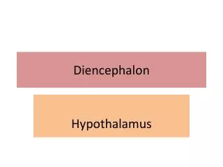
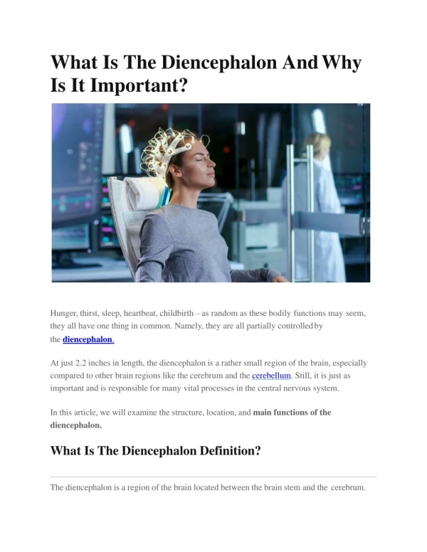
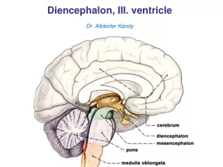
In the middle of the brain: the diencephalon. Diencephalon is a term used for a secondary brain vesicle during the development of the CNS. Those structures which arise from this are also summarized as diencephalon in the adult brain.
A brief summary of the early development of the CNS Note that the original cavity of the diencephalon becomes the third ventricle!
Medial and lateral geniculate bodies might be summarized as the methatalamus, but frequently they are considered as two nuclei of the thalamus.
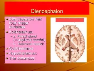
Parts of diencephalon and their main functions EPITHALAMUS striamedullaristhalami habenulartrigones habenularcomissure pinealgland posteriorcomissure THALAMUS SUBTHALAMUS subthalamicnucleus zonaincerta H-fields of Forel HYPOTHALAMUS METATHALAMUS medialgeniculate body, lateralgeniculatebody
Parts of diencephalon and their main functions EPITHALAMUS circadianrhythm (melatonin), limbicsystem THALAMUS relayingsensoryinformationtothecortex, some motor functions, regulation of consciousness SUBTHALAMUS regulation of movement, posture (extrapyramidalsystem) HYPOTHALAMUS endocrine and autonomic centre (METATHALAMUS) MGB: hearing, LGB: visualpathway
Appearance of diencephalon on different surfaces of the brain 1. Roof: choroid tela and plexus Floor: hypothalamus with optic and infundibular recesses Anterior: columns of fornix, anterior comissure, lamina terminalis, triangular recess Posterior: habenular and posterior comissures, suprapineal and pineal recesses Lateral: thalamus, hypothalamus corpus callosum septum pellucidum fornix ant. comissure pineal body lamina terminalis mesencephalon optic chiasm hypophysis pons triangularrecess opticrecess infundibularrecess suprapinealrecess pinealrecess Biggest proportion of the diencephalon is observable from the mediansagittal surface of the brain. Medial surface of the diencephalon gives the lateral wall of the third ventricle!
2. Roof of the third ventricle with the great cerebral vein formed by the fusion of the two internal cerebral veins. thalamus After lifting corpus callosum and the body of fornix, the top of the two thalami and between them the choroid thela giving the roof of the third ventricle is seen. Embedded into the choroid tela the two internal cerebral veins anteriorly and the great cerebral vein posteriorly become visible. corpus callosum with fornix
INTERVENTRICULAR FORAMEN taenia fornicis taenia choroidea stria medullaris taenia thalami choroid tela habenular triangle habenular comissure Line of attachment of the choroid tela to the stria medularis thalami, habenular trigones and habenular comissure is called taenia thalami. (Taenia thalami continues over the interventricular foramen in the lateral ventricle as the taenia choroidea).
corpus callosum LATERAL VENTR. LATERAL VENTR. fornix caudate nucleus * * thalamostriate vein in stria terminalis * * internal cerebral vein stria medullaris thalami (epithalamus) thalamus choroid lamina epithelialis: layer of modified ependymal cells persisting after thinning of the original wall of the brain vesicles III taenia: an artefact,line of attachment of the choroid tela (asterisks) hypothalamus choroid lamina epithelialis +pia mater = tela choroidea, and this together with the vascular network inside is the choroid plexus
optic chiasm 3. infundibulum, tuber cinereum optic tract mamilary bodies mesencephalon LGB Inferior portion of diencephalon reaches the basal surface of the brain. Note the interpeduncular fossa with the brainstem exit of the oculomotor nerve and the arterious circle of Willis enclosing basal aspect of the diencephalon: optic chiasm, infundibulum and mamillary bodies.
On the posterior pole of diencephalon medial and lateral geniculate bodies (together: metathalamus) and the pulvinar is directly seen, but pineal body (a part of epithalamus) is also easily found in the midline. Superior colliculus is connected to the LGB, while inferior colliculus projects into the MGB. 4. pulvinar LGB MGB Already done: • development of diencephalon • parts and their functions • contribution of diencephalon to various surfaces of the brain • walls and recesses of the third ventricle Diencephalon in coronal sections! posterior view of the brainstem and the diencephalon
corpus callosum sept. pellucidum internal capsule fornix • triangular recess • optic recess • infundibular recess optic tract (OT) HYPOTHALAMUS
corpus callosum sept. pellucidum fornix internal capsule OT EPITHALAMUS THALAMUS HYPOTHALAMUS
corpus callosum fornix internal capsule LGB MGB hippocampus pyramidal tract EPITHALAMUS THALAMUS SUBTHALAMUS METATHALAMUS