Download
1 / 88
890 likes | 908 Vues
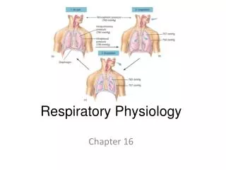
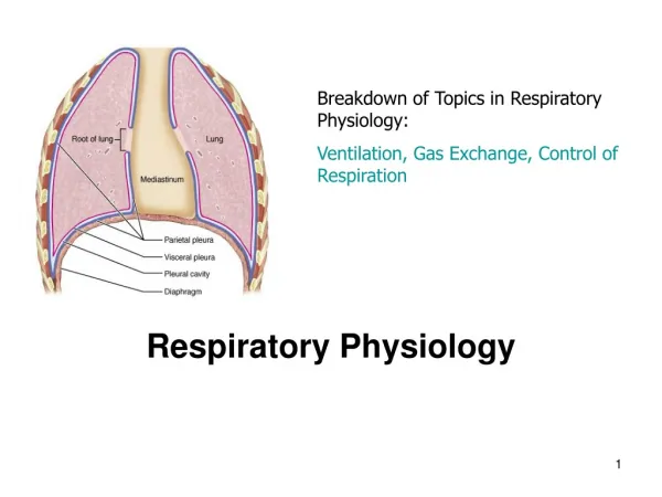
Respiratory Physiology. heart. heart. circuitry. heart. circuitry. tissues. lung. heart. circuitry. t issues. lung. heart. circuitry. t issues. cell. STRUCTURE of respiratory system Conducting zone Respiratory zone. STRUCTURE of respiratory system Conducting zone

E N D
heart circuitry
heart circuitry tissues
lung heart circuitry tissues
lung heart circuitry tissues cell
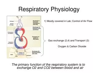
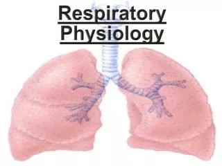
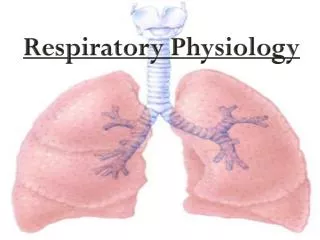
STRUCTURE of respiratory system • Conducting zone • Respiratory zone
STRUCTURE of respiratory system • Conducting zone • (nose, larynx, trachea, • bronchi, bronchioles) • The walls conducting airways • contain smooth muscle • Sympathetic • Parasympathetic • 2 receptors • relaxation, dilatation of the airway
STRUCTURE of respiratory system • Conducting zone • Anatomic dead space • V of the conducting airways • Physiologic dead space • V of the lungs that does not • participapate in gas exchance
STRUCTURE of respiratory system • Conducting zone • Physiologic dead space (VD) • V of the lungs that does not • participapate in gas exchance • PaCO2 – PECO2 • VD = VT x ---------------- • PaCO2 • VT – tidal volume • PaCO2 – PCO2 of arterial blood • PECO2 – PCO2 of mixed expired air
STRUCTURE of respiratory system Respiratory zone Alveoli Lung ~ 300 x 106 alveoli
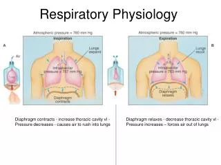
diaphragm inspiration expiration
inspiration – mm. intercosales externi expiration – mm. intercosteles interni
inspiration – mm. intercosales externi expiration – mm. intercosteles interni
inspiration – mm. intercosales externi expiration – mm. intercosteles interni
inspiration – mm. intercosales externi expiration – mm. intercosteles interni
inspiration – mm. intercosales externi expiration – mm. intercosteles interni
inspiration – mm. intercosales externi expiration – mm. intercosteles interni
pleura parietalis diaphragm inspiration expiration
pleura visceralis diaphragm inspiration expiration
pleura parietalis transmural pressure pleura visceralis diaphragm inspiration expiration
pleura parietalis pleura visceralis transpulmonal pressure diaphragm inspiration expiration
pleura parietalis pleura visceralis intrapulmonal pressure diaphragm inspiration expiration
pleura parietalis transmural pressure pleura visceralis intrapulmonal pressure transpulmonal p. diaphragm inspiration expiration
pleura parietalis transpulmonal p. pleura visceralis diaphragm inspiration expiration
PNEUMOTHORAX pleura parietalis transmural p. pleura visceralis bránice - diaphragma inspirace expirace
alveoli pleura parietalis transmural p. pleura visceralis intrapulmonal pressure transpulmonal p. diaphragm inspiration expiration
Physiologic dead space Is the volume of air in the lungs that does not participate in gas exchance (i.e.e it is dead) Anatomic dead space – is the volume of conducting airways Functional dead space volume – which is made up of alveoli that do not participate in gas exchance (alveoli that are ventilated, but are not perfused by pulmonary capilary blood)
Physiologic dead space PaCO2 – PECO2 V D = VT x ----------------------- PaCO2 VD – physiologic dead space (mL) VT – tidal volume (mL) PaCO2 – PCO2 of arterial blood (mmHg) PECO2 - PCO2 of expered air (mmHg)
Physiologic dead space PaCO2 – PECO2 V D = VT x ----------------------- PaCO2 40 - 30 VD = 500 x -------------- 40 = 500 x 0.25 = 125
Alveolar ventilation VA = (VT – VD) x breasths/min VA - alveolar ventilation (mL/min) VT - tidal volume (mL) VD - physiologic dead space (mL/min)
Alveolar ventilation VA = (VT – VD) x breasths/min VA = (500 – 125) x 16 = 375 x 16 = 6000 mL/min
lung heart circuitry tissues cell