Download
1 / 81
920 likes | 1.51k Vues
Respiratory physiology. Tom Archer, MD, MBA UCSD Anesthesia. www.argentour.com/tangoi.html. The dance of pulmonary physiology— Blood and oxygen coming together. But sometimes the match between blood and oxygen isn’t perfect!. http://www.bookmakersltd.com/art/edwards_art/3PrincessFrog.jpg.

E N D
Respiratory physiology Tom Archer, MD, MBA UCSD Anesthesia
www.argentour.com/tangoi.html The dance of pulmonary physiology— Blood and oxygen coming together.
But sometimes the match between blood and oxygen isn’t perfect! http://www.bookmakersltd.com/art/edwards_art/3PrincessFrog.jpg
Outline (1) • Failures of gas exchange • In anesthesia– think mechanical first! • Hypoxemia is easier to produce than hypercarbia—why? • Measuring severity of poor oxygenation • Two pulmonary players—the burly and weakling alveoli (V/Q mismatch) • Shunt • He3 MR imaging in V/Q mismatch • Diffusion barrier
Outline (2) Dead Space (anatomical + alveolar = physiologic) Capnography and ETCO2 Airway flow problems and flow volume loops Large airway-- Intra and extra thoracic Small airway (Intrathoracic, e.g. asthma, COPD) Pulmonary hypertension Exactly how does it kill patients? Interventricular septum bowing Common hemodynamic management of all stenotic cardiopulmonary lesions.
Failures of gas exchange Shunt Low V/Q Alveolar dead space Diffusion barrier High V/Q
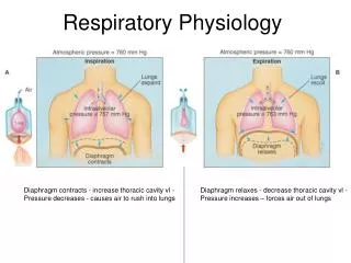
For gas exchange problems: • Always think of mechanical problems first: • Mainstem intubation • Partially plugged (blood, mucus) or kinked ETT. • Disconnect or other hypoventilation • Low FIO2 • Pneumothorax
For gas exchange problems: • Hand ventilate and feel the bag! • Examine the patient! • Look for JVD. • Do not Rx R mainstem intubation with albuterol! • Do not Rx narrowed ETT lumen with furosemide! • Consider FOB and / or suctioning ETT with NS. • THINK OF MECHANICAL PROBLEMS FIRST!
In life / medicine / gas exchange problems: • Beware of tunnel vision. Get used to asking yourself, “What am I not thinking of?” • “Asthma” = tracheal stenosis / tumor? • “Bronchospasm” = dried secretions in ETT? • Hypotension despite distended peripheral veins = pneumothorax? • “Coagulopathy” = chest tube in liver?
All That Wheezes Is Not Asthma: Diagnosing the Mimics www.mdchoice.com/emed/main.asp?template=0&pag...
Failures of gas exchangecausing hypoxemia • External compression of lung causing atelectasis. • Obesity, ascites, surgical packs, pleural effusion • Parenchymal disease (V/Q mismatch and shunt) • Asthma, COPD, pulmonary edema, ARDS, pneumonia, • Tumor, fibrosis, cirrhosis • (Intra-cardiac shunts)
Measuring severity of oxygenation problem: • A-a gradient (from alveolar gas equation). • Calculates “PAO2” • Needs FIO2, PB, PaCO2, PaO2 • Shunt fraction equation • Needs PAO2, CcO2, CvO2, CaO2 • PaO2 / FIO2 (< 200 in ARDS) • None of these give us etiology or physiology (shunt vs. V/Q mismatch).
Two pulmonary players: • The burly alveolus (high V/Q).
Two pulmonary players: • The weakling alveolus (low V/Q).
A fundamental question: • In terms of arterial O2 and CO2 tensions, can the burly alveolus compensate for the weaklingalveolus? • No, for PaO2. • Yes, for PaCO2. • This basic fact explains a lot. Know it cold.
Shunt, or “weakling” (low V/Q) alveolus SaO2 = 75% Normal alveolus SaO2 = 96% “Burly” (high V/Q) alveolus SaO2 = 99% Equal admixture of “weakling” and “burly” alveolar blood has SaO2 = (75 + 99)/ 2 = 87%. http://www.biotech.um.edu.mt/home_pages/chris/Respiration/oxygen4.htm Modified by Archer TL 2007
The weakling alveolus (shunt or V/Q mismatch) The burly alveolus pO2 = 50 mm Hg pO2 = 130 mm Hg SaO2 = 80% SaO2 = 98% SaO2 = 75% SaO2 = 75% pO2 = 50 mm Hg pO2 = 40 mm Hg pO2 = 130 mm Hg pO2 = 40 mm Hg Can the burly alveolus compensate for the weakling alveolus? Not for oxygen! The burly alveolus can’t saturate hemoglobin more than 100%. SaO2 of equal admixture of burly and weakling alveolar blood = 89%
Low V/Q alveoli cause widened A-a gradient, just like shunt Weakling Normal Burly http://www.lib.mcg.edu/edu/eshuphysio/program/section4/4ch5/s4ch5_11.htm
For CO2, burly alveolus CAN compensate for the weakling alveolus. Weakling alveolus Normal alveolus Burly alveolus Admixture of burly and weakling alveolar blood Average alveolar PACO2 = 40 mm Hg. Hence, PaCO2 = 40 mm Hg http://focosi.altervista.org/alveolarventilation2.jpg Modified by Archer TL
The weakling alveolus The burly alveolus pCO2 = 44 mm Hg pCO2 = 36 mm Hg pCO2 = 44 mm Hg pCO2 = 46 mm Hg pCO2 = 36 mm Hg pCO2 = 46 mm Hg Can the burly alveolus compensate for the weakling alveolus? Yes, for CO2! The burly alveolus, if it tries real hard, can blow off extra CO2. Pulmonary venous blood pCO2 and PaCO2 = 40 mm Hg
Shunt etiologies • Normal • Bronchial circulation • Thebesian veins • Intracardiac • Tetralogy of Fallot, VSD, etc. • Intrapulmonary • Bronchial intubation • Obesity • Cirrhosis • Osler-Weber-Rendu
Hypoxemia due to shunt • Increased FIO2 helps at low shunt percentages by dissolving more O2 in oxygenated blood. • At high shunt percentages, increased FIO2 does not help appreciably. • HPV decreases perfusion of hypoxic alveoli.
Normal shunt– bronchial circulation and Thebesian veins aorta Pulmonary veins http://www.lib.mcg.edu/edu/eshuphysio/program/section4/4ch5/s4ch5_10.htm Modified by Archer TL 2007
Intrapulmonary shunt in obesity: When FRC is below closing capacity, perfusion of non-ventilated alveoli is SHUNT.
V/Q mismatch • Emphasized by John West in the 1970’s. • Seen in most lung diseases. • Prototypes are: asthma, COPD, ARDS. • V/Q mismatch and shunt both cause hypoxemia despite possible hyperventilation (burly alveoli can’t compensate for weakling alveoli).
He3 MR showing ventilation defects in a normal subject and in increasingly severe asthmatics. Author Samee, S ; Altes T ; Powers P ; de Lange EE ; Knight-Scott J ; Rakes G Title Imaging the lungs in asthmatic patients by using hyperpolarized helium-3 magnetic resonance: assessment of response to methacholine and exercise challengeJournal Title Journal of Allergy & Clinical ImmunologyVolume 111 Issue 6 Date 2003 Pages: 1205-11
Baseline Methacholine Albuterol He3 MR scans – ventilation defects in asthmatics Modified by Archer TL 2007
Diffusion barrier (DB) to O2 and CO2and DLCO • Conceptually difficult • Thickened alveolar capillary membrane. • Exercise induced hypoxemia d/t dec transit time • DLCO related to many factors: • Membrane barrier thickness • Perfused alveolar surface area (COPD, lung resection) • Cardiac output • Hemoglobin concentration • DB not usually a significant clinical problem for us.
DLCO related to many factors: • Membrane barrier thickness • Perfused alveolar surface area (COPD, lung resection) • Cardiac output • Hemoglobin concentration http://www.lib.mcg.edu/edu/eshuphysio/program/section4/4ch3/s4ch3_25.htm
Diffusion in alveolar capillaries normally complete in 0.25 seconds. Thickened alveolar membrane may require more time for equilibration, which may not be available at higher cardiac outputs. Result: desaturation with exercise. http://www.lib.mcg.edu/edu/eshuphysio/program/section4/4ch3/s4ch3_27.htm
Dead space (DS) • Volume of expired gas which has not participated in gas exchange. • Physiological DS = Anatomical DS + Alveolar DS • VT (minute vent) = VA (alv vent) + VD (DS vent). • PaCO2 is inversely proportional to alveolar ventilation. • Know these facts cold.
PaCO2 is inversely proportional to alveolar ventilation. http://focosi.altervista.org/alveolarventilation2.jpg Modified by Archer TL
The same minute ventilation can cause markedly different amounts of alveolar ventilation, depending on tidal volume. http://www.lib.mcg.edu/edu/eshuphysio/program/section4/4ch3/s4ch3_22.htm
Anatomic and alveolar dead space • Anatomic dead space gas comes out BEFORE alveolar CO2. • Alveolar dead space gas comes out at the same time as CO2 from perfused alveoli. • Alveolar dead space gas DILUTES CO2 from perfused alveoli. This is why ETCO2 < PaCO2.
Capnographs– two types • CO2 vs. time (commonest, what we have). • CO2 vs. expired volume (more useful)
Anatomical dead space Single breath oxygen technique http://images.google.com/imgres?imgurl=http://www.lib.mcg.edu/edu/eshuphysio/program/section4/4ch3/4ch3img/page15b.jpg&imgrefurl=http://www.lib.mcg.edu/edu/eshuphysio/program/section4/4ch3/s4ch3_15.htm&h=379&w=271&sz=57&hl=en&start=33&tbnid=9bhXZpatrf-ajM:&tbnh=123&tbnw=88&prev=/images%3Fq%3Dalveolar%2Bventilation%2B%26start%3D20%26ndsp%3D20%26svnum%3D10%26hl%3Den%26lr%3D%26sa%3DN
http://images.google.com/imgres?imgurl=http://www.lib.mcg.edu/edu/eshuphysio/program/section4/4ch3/4ch3img/page15b.jpg&imgrefurl=http://www.lib.mcg.edu/edu/eshuphysio/program/section4/4ch3/s4ch3_15.htm&h=379&w=271&sz=57&hl=en&start=33&tbnid=9bhXZpatrf-ajM:&tbnh=123&tbnw=88&prev=/images%3Fq%3Dalveolar%2Bventilation%2B%26start%3D20%26ndsp%3D20%26svnum%3D10%26hl%3Den%26lr%3D%26sa%3DNhttp://images.google.com/imgres?imgurl=http://www.lib.mcg.edu/edu/eshuphysio/program/section4/4ch3/4ch3img/page15b.jpg&imgrefurl=http://www.lib.mcg.edu/edu/eshuphysio/program/section4/4ch3/s4ch3_15.htm&h=379&w=271&sz=57&hl=en&start=33&tbnid=9bhXZpatrf-ajM:&tbnh=123&tbnw=88&prev=/images%3Fq%3Dalveolar%2Bventilation%2B%26start%3D20%26ndsp%3D20%26svnum%3D10%26hl%3Den%26lr%3D%26sa%3DN www.lib.mcg.edu/.../section4/4ch3/s4ch3_15.htm
ETCO2 = 40 mm Hg With no alveolar dead space ETCO2 = 20 mm Hg With 50% alveolar dead space 20 40 40 20 Alveolar dead space gas (with no CO2) dilutes other alveolar gas. 0 40 40 40 46 0 46 46
Capnography • Obvious: picks up changes in ventilation (such as disconnection). • Not so obvious: picks up changes in pulmonary perfusion. • Commonest cause of abrupt fall in ETCO2 is hypotension (+ fall in PA pressure) with acute increase in alveolar dead space. • Also think air / clot embolus
Capnography • Upsloping alveolar plateau as sign of V/Q mismatch and / or delayed expiration. http://www.caep.ca/CMS/images/cjem/v53-169-f1.png
Diagnosing airway flow problems with flow volume loops. Clinically used and useful? Not! On the test? Probably! Interesting? Maybe.