Download
1 / 23
270 likes | 607 Vues
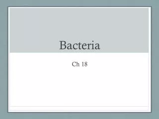
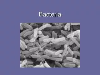
Bacteria. Bacteria. Prokaryotic Eukaryotic Can be Both. Prokaryotic! Bacteria are the ONLY prokaryotes on Earth!. Remember!. Prokaryotic vs. Eukaryotic Cells. Prokaryotic. Eukaryotic. Larger & more complex Contain a nucleus Contain membrane-bound organelles. Small & simple NO NUCLEUS

E N D
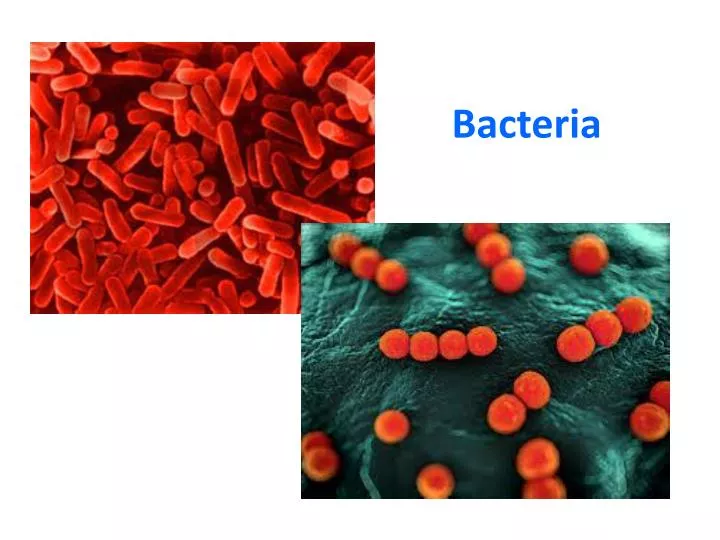
Bacteria • Prokaryotic • Eukaryotic • Can be Both Prokaryotic! Bacteria are the ONLY prokaryotes on Earth!
Prokaryotic vs. Eukaryotic Cells Prokaryotic Eukaryotic Larger & more complex Contain a nucleus Contain membrane-bound organelles • Small & simple • NO NUCLEUS • No membrane-bound organelles
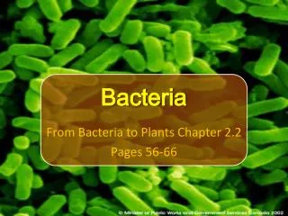
Bacteria • Unicellular • Multicellular • Can be Both Unicellular! Bacteria are composed of a single cell!
In what kingdoms can we find bacteria? • Protista • Fungi • Monera • None of the above Bacteria can be found in the kingdoms Archaebacteria & Eubacteria!
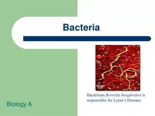
Archaebacteria vs. Eubacteria Archaebacteria Eubacteria Cell walls NOT made of peptidoglycan Live in extreme environments Cell walls made of peptidoglycan Identified using gram staining Prokaryotic Unicellular
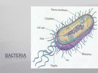
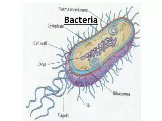
Structures of Bacterial Cells • Plasma Membrane: selectively permeable for exchange of nutrients and waste • Cytoplasm: fluid inside of the cell • Cell Wall
Structures of Bacterial Cells • DNA: free in the cytoplasm, but clustered in a “nucleoid region” • Flagella • Ribosomes
Structures of Bacterial Cells • Pili: small protein extensions used to anchor themselves OR to help exchange DNA with other bacteria
There are thousands of identified species of bacteria, but scientists have estimated that there are millions waiting to be discovered! So how do we classify all these bacteria? • Bacteria are classified according to: • Shape • Cell Wall • Metabolism
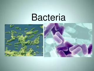
What are some differences between the 2 types of bacteria shown here? Shape! • Bacteria come in 3 shapes: • Round • Rod • Corkscrew
Classification by Shape • Coccus (cocci, pl.): round, can be found in clumps or lines • Baccillus (baccillis, pl.): rod shaped • Spirillum (spirilli, pl.): corkscrew (spiral) shaped
Spirillum Coccus Bacillus
Classification by Cell Wall • Eubacteria can be identified by the thickness of their cell wall. • Gram Staining determines how thick the cell wall is. • Gram + Bacteria: stains purple, have a thick cell wall • Gram - Bacteria: stains pink, have a thin cell wall Gram Negative (-) Gram Positive (+)
Gram + • Gram + • Gram – • Both • Neither Bacillus anthracis
Gram - • Gram + • Gram – • Both • Neither Spirillumvolutans
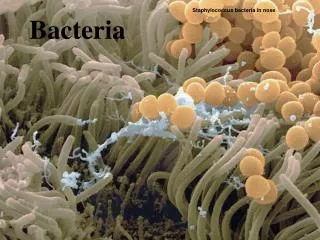
Gram + and Gram - • Gram + • Gram – • Both • Neither Staphylococcus aureus& Escherichia coli
A scientist uses gram staining on a colony of bacteria that she collected from a volcanic vent. What will her results be? Neither! The bacteria collected was archaebacteria. Gram staining will be ineffective, because archaebacteria do not have peptidoglycan in their cell walls. Gram staining can only be used to identify eubacteria! • Gram + • Gram – • Both • Neither
Classification By Metabolism • Classified by how they obtain energy • Can be heterotrophic – obtains food from another source • Can be autotrophic – makes own food • Chemoautotrophs: make own food with inorganic molecules • Photoautotrophs: use photosynthesis
Where can we find bacteria? What do you think the dirtiest part of the classroom is? • Your group will swab a location of your choice & see if bacteria are present. • We will check the growth in a few days.
Directions to Prepare the Petri Dish • Pre-Lab Questions • Write your group’s info on the bottom (initials of all of the members, & class period) • Agree on a location to test. Come to me for a sterile Q-tip. • Rub 1 side of the Q-tip on the location, then lightly rub the same side onto the agar. • Draw your dish on your handout. • Replace the lid & tape it shut. • Place it on the tray.