Download
1 / 35
380 likes | 583 Vues
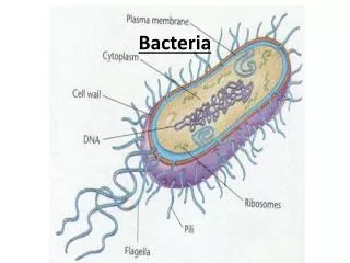
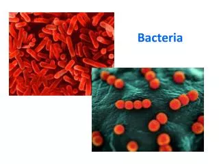
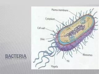
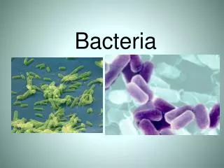
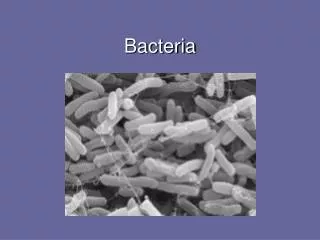
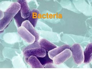
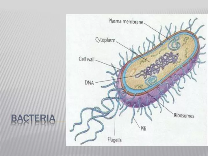
Bacteria. Characteristics. Prokaryotes Microscopic (Eukaryotic cells are at least 10x bigger) Unicellular DNA is a single circular piece of DNA Asexual Reproduction Binary Fission Metabolism Aerobic Anaerobic. Genetic Exchange Conjugation –transfer DNA through contact

E N D
Characteristics • Prokaryotes • Microscopic (Eukaryotic cells are at least 10x bigger) • Unicellular • DNA is a single circular piece of DNA • Asexual Reproduction • Binary Fission • Metabolism • Aerobic • Anaerobic
Genetic Exchange • Conjugation –transfer DNA through contact • Transformation – acquire DNA from dead bacteria • Transduction – DNA is transferred from one bacteria to another using a virus (genetic engineering)
http://highered.mcgraw-hill.com/sites/0072556781/student_view0/chapter13/animation_quiz_2.htmlhttp://highered.mcgraw-hill.com/sites/0072556781/student_view0/chapter13/animation_quiz_2.html http://highered.mcgraw-hill.com/sites/0072556781/student_view0/chapter13/animation_quiz_3.html
Survival of the Fittest!!! Bacteria have been around for 3.5 billion years!! How???? • Cell Walls • Capsules (surrounds cell wall) • Asexual Reproduction, but can still acquire other genes • Inhabit every place on Earth
allow them to withstand drought, high temps., lack of food, etc. endospores
Shapes • Coccus : Spheres • Bacillus : Rods • Spirillum : Spirals • Arrangements • Strept : Chains • Staph : Clusters • Diplo : Pairs Bacteria are Classified according to Shape and arrangement of cells
Gram Stain (pg. 529) • Gram + simple walls, large amount of peptidoglycan • Gram - less peptidoglycan, outer membrane contains lipopolysaccharides which are often toxic and provides additional protection more resistant to antibiotics • Many antibiotics (penicillens) inhibit synthesis of cross links in peptidoglycan and prevent formation of a functional wall Gram negative Gram positive
Gram Positive Organisms • Aerobic, Gram-positive cocci • Staphylococcus aureus (fig 1, 2, 3, 4) • Staphylococcus epidermidis (fig 1) • Staphylococcus sp. (Coagulase-negative)(fig 1) • Streptococcus pneumoniae (Viridans group)(fig 1, 2, 3) • Streptococcus agalactiae (group B)(fig 1) • Streptococcus pyogenes (group A)(fig 1, 2) • Enterococcus sp.(fig 1, 2, 3) • Aerobic, Gram-positive rods • Bacillus anthracis (fig 1, 2) • Bacillus cereus (fig 1, 2) • Bifidobacterium bifidum (fig 1) • Lactobacillus sp. (fig 1, 2) • Listeria monocytogenes (fig 1, 2) • Nocardia sp.(fig 1, 2) • Rhodococcus equi (coccobacillus)(fig 1) • Erysipelothrix rhusiopathiae (fig 1) • Corynebacterium diptheriae (fig 1, 2) • Propionibacterium acnes (fig 1) • Anaerobic, Gram-positive rods • Actinomyces sp. (fig 1, 2) • Clostridium botulinum (fig 1) • Clostridium difficile (fig 1) • Clostridium perfringens (fig 1, 2, 3) • Clostridium tetani (fig 1, 2) • Anaerobic, Gram-positive cocci • Peptostreptococcus sp. (fig 1)
Gram Negative Organisms • Aerobic, Gram-negative cocci • Neisseria gonorrhoeae (fig 1, 2, 3, 4) • Neisseria meningitidis (fig 1; false color of the bacterium., 2) • Moraxella catarrhalis (fig 1) • Anaerobic, Gram-negative cocci • Veillonella sp. (fig 1) • Aerobic, Gram-negative rods • Fastidious, Gram-negative rods • Actinobacillus actinomycetemcomitans (fig 1) • Acinetobacter baumannii(fig 1 really A. calcoaceticus) • Bordetella pertussis (fig 1, 2) • Brucella sp. (fig 1) • Campylobacter sp.(fig 1) • Capnocytophaga sp.(fig 1,2) • Cardiobacterium hominis (fig 1) • Eikenella corrodens (fig 1) • Francisella tularensis (fig 1,) • Haemophilus ducreyi (fig1,2) • Haemophilus influenzae (fig 1, 2) • Helicobacter pylori (fig 1, 2, 3, 4) • Kingella kingae (fig ) • Legionella pneumophila (fig 1, 2, 3) • Pasteurella multocida (fig 1) • Enterobacteriaceae (glucose-fermenting Gram-negative rods) • Citrobacter sp. (fig 1) • Enterobacter sp. (fig 1) • Escherichia coli (fig 1, 2) • Klebsiella pneumoniae (fig1, 2) • Proteus sp. (fig 1) • Salmonella enteriditis (fig 1) • Salmonella typhi (fig 1) • Serratia marcescens (fig 1, 2) • Shigella sp. (fig 1) • Yersinia enterocolitica (fig 1) • Yersinia pestis (fig 1, 2) • Oxidase-positive, glucose-fermenting Gram-negative rods • Aeromonas sp. (fig 1) • Plesiomonas shigelloides (fig 1) • Vibrio cholerae (fig 1, 2) • Vibrio parahaemolyticus (fig 1) • Vibrio vulnificus (fig 1) • Glucose-nonfermenting, Gram-negative rods • Acinetobacter sp. (fig 1) • Flavobacterium sp. (fig 1) • Pseudomonas aeruginosa (fig 1, 2) • Burkholderia cepacia (fig 1) • Burkholderia pseudomallei (fig 1) • Xanthomonas maltophilia or Stenotrophomonas maltophila(fig 1) • Anaerobic, Gram-negative rods • Bacteroides fragilis (fig 1) • Bacteroides sp. (fig 1) • Prevotella sp. (fig 1) • Fusobacterium sp. (fig 1,2) • Gram-negative spiral • Spirillum minus (minor)- (fig 1)
Nutrition • Autotrophic • Photosynthetic • Chemoautotrophic (nitrogen fixers) • Heterotrophic • Decomposer • Parasitic (Treponema pallidum)
Important Recyclers in environment • Nitrogen cycle
Bacteria can produce chemicals • Acetone, Butanol
Bacteria are used to make food • Pickles, buttermilk, cheese, sauerkraut, olives, vinegar, sourdough bread, beer, wine
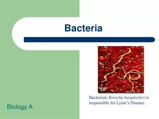
Bacteria cause disease • Produce toxins (Clostridium botulinum) • Metabolize their host (Mycobacterium tuberculosis)
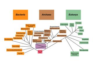
History of Microbiology • 1664: Robert Hooke - microscope • 1684: Antoni van Leeuwenhoek - microorganisms • 1798: Edward Jenner - smallpox vaccination • 1864: Louis Pasteur - spontaneous generation • 1884: Robert Koch - Koch’s postulates • 1889: Martinus Beijerink - concept of virus • 1929: Alexander Fleming - discovery of penicillin • 1977: Carl Woese - discovery of Archaea • 1981: First reports of AIDS • 1983: Luc Montagnier - discovery of HIV • 1995: Craig Venter - complete genome sequence