Download
1 / 54
570 likes | 1.14k Vues
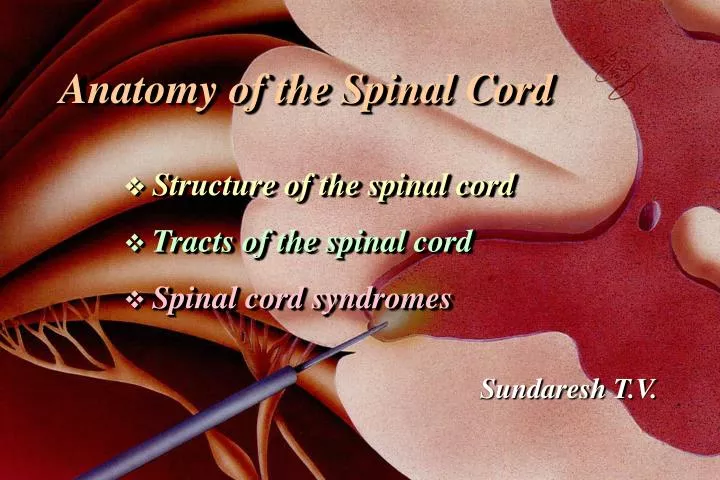
Anatomy of the Spinal Cord Structure of the spinal cord Tracts of the spinal cord Spinal cord syndromes. Sundaresh T.V. Spinal Cord. - Comparable to Input-Output (IO) System of the Computer - Spinal Nerve (C8, T12, L5, S5, Cx1) - Segmental Structure of Neural Tube Origin.

E N D
Anatomy of the Spinal Cord • Structure of the spinal cord • Tracts of the spinal cord • Spinal cord syndromes Sundaresh T.V.
Spinal Cord - Comparable to Input-Output (IO) System of the Computer - Spinal Nerve (C8, T12, L5, S5, Cx1) - Segmental Structure of Neural Tube Origin
Spinal segment C8, T12, L5, S5, Cx1 Anterior (Ventral) Root Posterior (Dorsal) Root Dorsal Root (Spinal) Ganglion Root - Rootlets
Spinal Cord External Figure Conus Medullaris (L1-2) Spinomedullary Junction - Foramen Magnum, Pyramidal decussation, C1 ventral root Enlargements - cervical (C5-T1) & lumbosacral (L1-L4) Longitudinal Fissures - anterior median fissure - anterolateral fissure - posterior median sulcus - posterolateral sulcus
Conus Medullaris (L1-2) Cauda Equina Anterior median fissure Anterolateral fissure
Posterior median sulcus Posterolateral sulcus Posterior intermediate sulcus Fasciculus cuneatus Fasciculus gracilis
Spinal Cord Meninges Periosteum of Vertebra - Epidural Space ----------------- epidural anesthesia Dura Mater Spinalis Arachnoid Membrane - Subarachnoid Space -------- Lumbar Puncture Spinal Anesthesia Pia Mater Spinalis - Denticulate Ligament --------- Cordotomy - Filum Terminale
Meninges of • the spinal cord • Dura mater • Arachnoid membrane • Pia mater Denticulate ligament - specilization of the pia mater - landmark for cordotomy
Spinal Cord Vascular Supply Arterial Supply - Spinal Arteries Anterior (1) & Posterior (2) Spinal Artery from Vertebral artery - Radicular Arteries ----- Segmental arteries from Vertebral, Ascending Cervical, Intercostal and Lumbar Artery Venous Drainage - Longitudinal & Radicular Veins to Intervertebral veins ---- to Internal Vertebral Venous Plexus to external vertebral venous plexus ---- to segmental veins
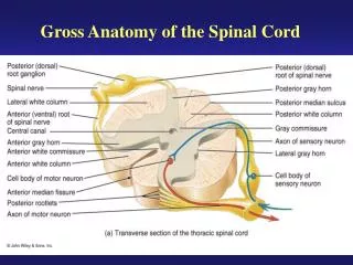
Spinal Cord Internal Structure White Matter Anterior Funiculus (Anterior White Column) Posterior Funiculus (Posterior White Column) Fasciculus Gracilis & Fasciculus Cuneatus Lateral Funiculus (Lateral White Column) Gray Matter Anterior Horn ------------ --- motor Posterior Horn -------------- sensory Lateral Horn ----------------- autonomic (sympathetic) Gray Commissure -------- anterior and posterior
1. posterior horn 2. anterior horn 3. intermediate zone (intermediate gray) 4. lateral horn 5. posterior funiculus 6. anterior funiculus 7. lateral funiculus 8. Lissauer's tract 9. anterior median fissure 10. posterior median sulcus 11. anterolateral sulcus 12. posterolateral sulcus 13. Posterior intermediate sulcus
cervical enlargement (C8) thoracic cord (T8) lumbal enlargement (L3) sacral cord (S1)
Spinal Cord Internal Structure Principles of Cord Organization 1) Longitudinal Arrangement Fibers (White Matter) ------------- White Column Cell Groups (Gray Matter) ------- Gray Column 2) Transverse Arrangement Afferent & Efferent Fibers Crossing (Commissural and Decussating) Fibers 3) Somatotopical Arrangement
Spinal CordInternal Structure Lamina of Rexed Lamina I ---------- posteromarginal nucleus Lamina II ---------- substantia gelatinosa of Rolando Lamina III, IV ----- nucleus proprius Lamina V, VI Lamina VII --------- intermediate gray intermediolateral cell column (ILM) Clarke’s column (Nucleus dorsalis) intermediomedial cell column (IMM) Lamina VIII Lamina IX ---------- anterior horn (motor) cell Lamina X ----------- gray commissure
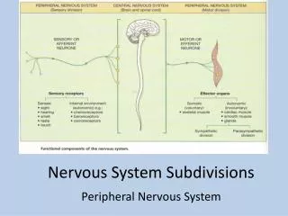
Spinal Cord Tracts Ascending Tracts Modality: Touch, Pain, Temperature, Kinesthesia Receptor: Exteroceptor, Interoceptor, Proprioceptor Primary Neuron: Dorsal Root Ganglion (Spinal Ganglion) Secondary Neuron: Spinal Cord or Brain Stem (Tertiary Neuron): Thalamus (Ventrobasal Nuclear Complex) Termination: Cerebral Cortex, Cerebellar Cortex, or Brain Stem
Spinal Cord Tracts Ascending Tracts Posterior White Column-Medial Lemniscal Pathway Spinothalamic Tract Spinoreticular or Spinoreticulothalamic Tract Spinocerebellar Tract Spinomedullothalamic Tract Cervicothalamic or Spinocervicothalamic Tract Spino-olivary Tract Spinotectal Tract
Spinal Cord Ascending Tracts Posterior White Column-Medial Lemniscal Pathway Modality: Discriminative Touch Sensation (include Vibration) and Conscious Proprioception (Position Sensation, Kinesthesia) Receptor: Most receptors except free nerve endings Ist Neuron: Dorsal Root Ganglion (Spinal Ganglion) Posterior Root - Posterior White Column 2nd Neuron: Dorsal Column Nuclei(Nucleus Gracilis et Cuneatus) Internal Arcuate Fiber - Lemniscal Decussation - Medial Lemniscus 3rd Neuron: Thalamus (VPLc) Internal Capsule ----- Corona Radiata Termination: Primary Somesthetic Area (S I)
medial lemniscus lemniscal decussation internal arcuate fiber posterior white column posterior root Posterior White Column - Medial Lemniscal Pathway - ipsilateralloss of discriminative touch sensation and conscious proprioception belowthe level of lesion
Spinal Cord Ascending Tracts Spinothalamic Tract Modality: Pain & Temperature Sensation, Light Touch Receptor: Free Nerve Ending Ist Neuron: Dorsal Root Ganglion (Spinal Ganglion) Posterior Root 2nd Neuron: Dorsal Horn (Lamina I, IV, V) Spinothalamic Tract - (Spinal Lemniscus) 3rd Neuron: Thalamus (VPLc, CL & POm) Internal Capsule ----- Corona Radiata Termination: Primary Somesthetic Area (S I) & Diffuse Widespread Cortical Region
spinothalamic tract anterior white commissure posterior root decussation Spinothalamic Tract - contralateral loss of pain and temperature sensation below the level of lesion
NeoSTTPaleoSTT Primary Motor Area (M I) VPLc (ventrobasal nuclear complex) (spinal lemniscus) spinothalamic tract Widespread cortical region CL (intralaminar thalamic nuclei) reticulothalamic pathways spinoreticular tract thalamus reticular formation Spinothalamic Tract & Spinoreticular Tract
Comparison of Fast and Slow Pain------ Spinothalamic Tract Fast PainSlow Pain Sharp, pricking Dull, burning Group III (A) fiber Group IV (C) fiber Short latency Slower onset Well localized Diffuse Short duration Long duration Less emotional Emotional, autonomic response Not blocked by morphine Blocked by morphine Neospinothalamic Tract Paleospinothalamic Tract
Spinal Cord Ascending Tracts Spinocerebellar Tract Modality: Unconscious Proprioception Receptor: Muscle spindle, Golgi tendon organ Ist Neuron: Dorsal Root Ganglion (Spinal Ganglion) Posterior Root , [Posterior Column] 2nd Neuron: 1. Clarke’s column Posterior Spinocerebellar Tract 2. Accessory Cuneate Nucleus Cuneocerebellar Tract 3. Posterior Horn Anterior Spinocerebellar r Tract Termination: Cerebellar Cortex
Anterior Spcbltr (superior cerebellar peduncle) anterior spinocerebellar tract anterior white commissure posterior root Posterior SpCblTr Inferior cerebellar peduncle cuneocerebellar tract (upper body) posterior white column posterior root Inferior cerebellar peduncle posterior spinocerebellar tract Clarke’s column posterior white column posterior root Spinocerebellar Tract
Spinal Cord Descending Tracts Corticospinal Tract Origin: Cerebral Cortex Brodmann Area 4 (Primary Motor Area, M I) Brodmann Area 6 (Premotor Area, PM ) Brodmann Area 3,1,2 (Primary Somesthetic Area, S I) Brodmann Area 5 (Anterior Portion of Sup. Parietal Lobule) Corona Radiata lnternal Capsule, Posterior Limb Crus Cerebri, Middle Portion Longitudinal Pontine Fiber Pyramid - pyramidal decussation Corticospinal Tract - Lateral and Anterior Termination: Spinal Gray (Rexed IV-IX)
Corona Radiata lnternal Capsule, Posterior Limb Crus Cerebri, Middle Portion Longitudinal Pontine Fiber Pyramid Pyramidal Decussation Corticospinal Tract - Lateral and Anterior CR IC LPF Corticospinal Tract Pyr LCST PD - ipsilateralUMN syndrome at the level of lesion ACST
Spinal Cord Descending Tracts Descending Tracts from Brain Stem Dorsolateral (Motor) Pathway Rubrospinal Tract Ventromedial (Motor) Pathway Tectospinal Tract Vestibulospinal Tract MLF (Medial Longitudinal Fasciculus) - interstitiospinal tract Sensory Modulation pathways Raphespinal & Cerulospinal Pathways Descending Autonomic Pathways
Spinal Cord Tracts ventromedial pathway dorolateral pathway Descending Tracts from Brain Stem
SOMATIC MOTOR SYSTEM upper motor neuron UMN Brain Stem Descending Pathway Rubrospinal Tract Tectospinal Tract Vestibulospinal Tract MLF Reticulospinal Tract Final Common Pathway VOLUNTARY CONTROL lower motor neuron LMN Pyramidal Tract AUTOMATIC CONTROL REFLEX EFFECTORS skeletal muscle
Spinal Cord Syndrome Location of Symptoms in Spinal Disease ipsilateral to lesion contralateralto lesion
Upper Motor Neuron (UMN) vs Lower Motor Neuron (LMN) Syndrome UMN syndromeLMN Syndrome Type of Paralysis Spastic Paresis Flaccid Paralysis Atrophy No (Disuse) Atrophy Severe Atrophy Deep Tendon Reflex Increase Absent DTR Pathological Reflex Positive Babinski Sign Absent Superficial Reflex AbsentPresent Fasciculation and Absent Could be Fibrillation Present
Spinal Cord Syndrome • Predominantly Motor Syndromes • Poliomyelitis (Infantile Paralysis) • - viral infection of lower motor neuron • - LMN syndrome at the level of lesion • Amyotrophic Lateral Sclerosis (ALS) • - combined LMN and UMN lesion • - LMN syndrome at the level of lesion • - UMN syndrome below the level of lesion • - Lou Gehrig’s disease in USA
Spinal Cord Syndrome 1. corticospinal tract 2. lower motor neuron (LMN) Amyotrophic Lateral Sclerosis
Spinal Cord Syndrome Amyotrophic Lateral Sclerosis (ALS) Lou Gherig’s Disease Lou "The Iron Horse" Gehrig (1903-41) 3.40, 2131(1925-39), 23 GSH, 147 RBI avg.
Spinal Cord Syndrome Amyotrophic Lateral Sclerosis (ALS) Lou Gherig’s Disease Stephen Haking (1946- ) British Physicist, A Brif History of Time
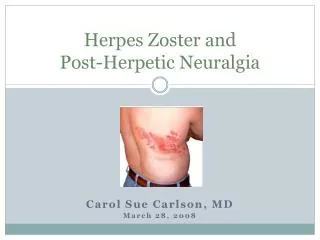
Spinal Cord Syndrome • Predominantly Sensory Syndromes • Herpes Zoster • - inflammatory reactions of spinal ganglion • - severe pain on the dermatomes of affected ganglion • Tabes Dorsalis • - common variety of neurosyphilis • - posterior colum and spinal posterior root lesion • - loss of discriminative touch sensation and conscious • proprioception below the level of lesion • - posterior column ataxia • - lancinating pain • - loss of deep tendon reflex (DTR)
Herpes Zoster (Shingles) • varicella-zoster virus • reactivation from • the dorsal root ganglia • unilateral vesicular • eruption within • a dermatome • T3 to L3 dermatome • lesions are frequent • zoster ophtahalmicus • (ophthalmic division • of trigeminal n., V1) • Ramsey-Hunt syndrome • (sensory br. of VII) • acyclovir, antiviral agent
Spinal Cord Syndrome Subacute Combined Degeneration (Combined System Disease) Lesion - posterior white column - corticospinal tract (UMN) Symptom - loss of discriminative touch sensation and conscious proprioception below the level of lesion - ipsilateral UMN syndrome below the level of lesion
Spinal Cord Syndrome 1. corticospinal tract 2. posterior white column Subacute Combined Degeneration