Download
1 / 19
190 likes | 465 Vues
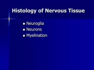
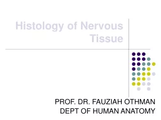
Histology Slides for Nervous Tissues. Slides are presented in order of magnification if different view are presented. As you view the following slides make sure you can accomplish these goals: Can you identify the tissue observable on the slides?

E N D
Histology Slides for Nervous Tissues • Slides are presented in order of magnification if different view are presented. • As you view the following slides make sure you can accomplish these goals: • Can you identify the tissue observable on the slides? • Can you identify the specific structures or layers indicates by the numbered arrows or brackets? • At the end of a sequence you will find the answers to the above for each slide.
1 1 1 1 3 2 2 3 100X
3 3 2 1 2 4 400X
5 5 5 5 400X
8 8 7 7 8 8 6 100X
7 8 6 11 9 10 400X
8 9 11 10 12 12 7 400X
Answers to Slides 2 through 7 Slide 2: Smear of gray matter low power. Slide 3: Smear of gray matter high power. Slide 4: Longitudinal view of myelinated nerve fibers. Slide 5: X-section of a nerve low power (Masson stain) Slide 6: X-section of a nerve high power (Masson stain) Slide 7: X-section of a nerve high power (H&E stain) Answers to the numbers: • Multipolar neuron cell body with nissl bodies, nucleus, and nucleolus. • Nerve fibers (axons of dendrites) • Neuroglial nuclei • Nucleus and nucleolus • Node of Ranvier • Epineurium
Answers to Slides 2 through 7 Answers to the numbers: • Perineurium • Nerve fasiscle • Endoneurium • Myelin sheath • Nerve fiber • Nucleus of Schwann cells
5 4 3 2 1 100X
5 4 3 2 1 400X
7 10 7 10 15 14 9 6 9 6 13 16 8 8 11 11 12 100X
11 5 17 100X 18
11 17 5 19 20 400X
11 18 17 17 17 400X
5 19 20 100X
23 22 21 21 21 24 100X
Answers to Slides 10 through 17 Slide 10 - 16: X-section of spinal cord Slide 17: Section through Dorsal Root Ganglion Answers to the numbers: • Dura mater. • Arachnoid mater. • Subarachnoid space. • Pia mater. • X-section through white matter. • Gray matter • Posterior or dorsal white column • Anterior or ventral white column • Lateral white column • Doral gray horn • Ventral gray horn
Answers to Slides 10 through 17 Answers to the numbers: • Anterior or ventral median fissure • Central canal • Posterior gray commissure • Posterior or dorsal median sulcus • Anterior gray commissure • Multipolar neuron cell body • Transverse nerve fibers • X-section through a nerve fiber • Myelin sheath created by Oligodendrocytes • Sensory neuron cell bodies • Longitudinal view of myelinated nerve fibers • Nodes of Ranvier • Satellite cells