Download
1 / 42
460 likes | 909 Vues
EPITHELIAL TISSUES. You’d be lost (dead) without them. WHAT ARE EPITHELIA ?. A. simple definition - "a layer of cells with a free surface". B. better definition - single or multiple layers of cells characterized by,. 1. a layer or layers composed of closely aggregated, polyhedral cells.

E N D
EPITHELIAL TISSUES You’d be lost (dead) without them.
WHAT ARE EPITHELIA? A. simple definition - "a layer of cells with a free surface" B. better definition -single or multiple layers of cells characterized by, 1. a layer or layers composed of closely aggregated, polyhedral cells 2. one side of cell layer(s) has a free surface 3. little intercellular substance between cells 4. cells cohere (stick) strongly to each other 5. cells form a sheet that covers a surface
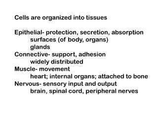
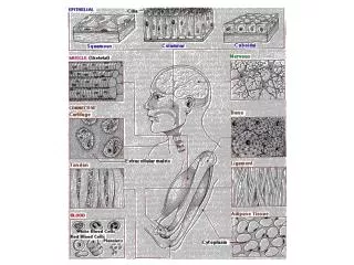
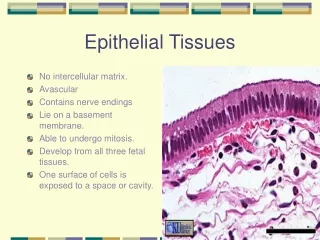
Functions of Epithelia In embryonic terms we can say that epithelia are derived from all 3 major germ layers, i.e. ectoderm, mesoderm, endoderm. A. Covering and lining surfaces (a barrier) Examples: a. skin b. epithelial cells (endothelium) lining blood vessels, c. mesothelium of peritoneal cavity (coelom)
FUNCTIONS OF EPITHELIA B. Regulation of materials and sensory information passing between, into, or out of organs/tissues 1. absorption (e.g. tall columnar epithelium of intestine) 2. secretion (e.g. epithelial lining of glands) 3. sensation (e.g. sensory cells, neuroepithelium - taste buds) 4. lubrication (e.g. mucus secreting epithelium of digestive tract) C. In some cases contractility (e.g. myoepithelium - often associated with glands such as sweat and mammary glands) D. Protection from the external environment
BASIC CELL SHAPES FOUND IN EPITHELIA A.squamous- flat - very thin (flat) in cross-section B.cuboidal- appear square in cross-section C.columnar- appear rectangular (tall, column shaped) in cross-section
1. NOTE THAT HOW AN EPITHLIUM LOOKS IN HISTOLOGICAL SECTIONS DEPENDS ON THE ORIENTATION OF THE EPITHELIUM WHEN SECTIONS ARE CUT. A CUBOIDAL EPITHELIUM CAN LOOK COLUMNAR, ETC. 2. ALSO NOTE THAT THE THREE SHAPES GIVEN FOR EPITHELIA ARE DERIVED FROM THEIR SHAPE AS THEY ARE SEEN IN HISTOLOGICAL CROSS-SECTIONS. IN REALITY, THE CELLS ARE REALLY POLYHEDRAL (MANY SIDED) IN THREE DIMENSIONS
3. There are also intermediate shapes of some epithelial cell types that lie between the major types described above. 4. The shape of the nucleus often corresponds to cell shape. Squamous epithelial cells, nucleus flattened - long axis parallel to basement membrane Cuboidal epithelial cells, nucleus - spherical Columnar - nucleus oblong, long axis perpendicular to the basement membrane 5. The shape of the nucleus is sometimes important in distinguishing different types of epithelia since often the cell borders are indistinct despite staining.
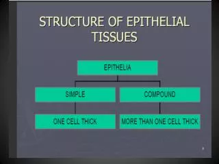
CLASSIFICATION OF EPITHELIA A. Simple epithelia - epithelia consisting of one layer of cells 1. Simple squamous - one layer of flattened cells covering a surface, e.g. endothelium of blood vessels. 2. Simple cuboidal - one layer of cuboidal cells covering a surface, e.g. epithelial lining of proximal and distal convoluted tubules in kidney. 3. Simple columnar - one layer of columnar cells covering a surface, e.g. epithelial lining of small intestine. a. Simple “low” columnar b. Simple “tall” columnar
B. Stratified epithelial types- more than one layer 1. keratinized, stratified, squamous epithelium - e.g. skin. Actually, usually only the cells at or near the free surface are squamous. Deeper cells tend to be cuboidal, columnar or polyhedral. 2. non-keratinized, stratified squamous (mucosal) epithelium - e.g. line wet cavities like the mouth, nasal passages and vagina. This epithelium often (but not always) has glands associated with it. As above, only the cells at or near the free surface are squamous. 3. stratified columnar epithelium - rare - e.g. parts of urethra, some ducts of the paratid gland. Only the cells at the free surface are columnar. 4. stratified cuboidal epithelium - rare, found in some glandular ducts e.g. sweat glands, sub-maxillary glands. 5. transitional epithelium - intermediate form of cells. Cells are often binucleate, cells at free surface may bulge into adjacent lumen, e.g. inner lining of urinary bladder. 6. pseudostratified epithelium - Looks stratified due to varying positions of cell nuclei, but all cells are in contact with the basement membrane, e.g. ciliated, pseudostratified, columnar epithelium of respiratory tract.
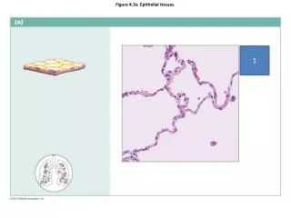
Types of epithelia - examples Kidney - Bowman’s capsule http://www.echt.chm.msu.edu/courseware/blockII/Pathology/RenalArchitecture_2.html
Types of epithelia - examples http://www.lab.anhb.uwa.edu.au/mb140/CorePages/Epithelia/Epithel.htm#labduo
Types of epithelia - examples Cornea Non-keratinized http://www.anatomy.dal.ca/html/Human%20Histology/DHD/Lab2/Laboratory2.html Inner lip epithelium http://www.anatomy.dal.ca/html/Human%20Histology/DHD/Lab2/Laboratory2.html http://www.lab.anhb.uwa.edu.au/mb140/CorePages/Epithelia/Epithel.htm#labduo
Types of epithelia - examples Keratinized http://www.lab.anhb.uwa.edu.au/mb140/CorePages/Integumentary/Integum.htm http://www.mhhe.com/biosci/ap/histology_mh/stratepi.html
Types of epithelia - examples www.mhhe.com/biosci/ ap/histology_mh/stratcuc.html Glandular duct http://hist.class.kmu.edu.tw/Basic/Epithelial/Stratified/Cuboidal/ep2c3h48_2.htm
Types of epithelia - examples http://www.lab.anhb.uwa.edu.au/mb140/CorePages/Epithelia/Epithel.htm#labduo
Types of epithelia - examples http://www.lab.anhb.uwa.edu.au/mb140/CorePages/Epithelia/Epithel.htm#labduo
Types of epithelia - examples http://www.lab.anhb.uwa.edu.au/mb140/CorePages/Epithelia/Epithel.htm#labduo http://www.udel.edu/Biology/Wags/histopage/colorpage/cep/ceptd.GIF
GENERAL CHARACTERISTICS OF EPITHELIA AND WHY EPITHELIA HAVE THESE CHARACTERISTICS I. The basement membrane is visible with the light microscope, particularly when silver impregnation or the PAS (Periodic Acid Schiff stain - stains carbohydrates) methods are used. Staining properties are due to the basement membrane having a high glycoprotein content. The basement membrane stains dark purple in the preparation of kidney tubules below. http://www.mc.vanderbilt.edu/histo/BasicTissue/Epith.char.html
GENERAL CHARACTERISTICS OF EPITHELIA AND WHY EPITHELIA HAVE THESE CHARACTERISTICS There is more to the basement membrane than just a layer of glycoprotein. A. When examined with the electron microscope, an epithelium has a basal lamina (50 - 80 nm, not resolved by light microscope) 1. Found wherever an epithelium contacts connective tissue. Visible only at EM level. Consists of granular layer of thin fibrils that is composed of a. collagen, b. a glycoprotein called laminin, and c. other large molecules called proteoglycans. 2. Forms a partial barrier (semipermeable) to diffusion between epithelium and underlying tissue (usually connective tissue). 3. Evidence suggests that the basal lamina is formed by the epithelial cells rather than the connective tissue. http://www.med.uiuc.edu/histo/small/atlas/image/tem38a/16250a1.htm
GENERAL CHARACTERISTICS OF EPITHELIA B. Basal lamina consists of the, *lamina densa - a fibrous, darkly staining layer into which fibers from hemidesmosomes, as well as adjacent extracellular matrix, insert * lamina rara (lamina lucida, reticular lamina) - a clear, non-staining layer (may be on one or both sides of the lamina densa). http://www.med.uiuc.edu/histo/large/atlas/image/tem38a/48750a1.htm
GENERAL CHARACTERISTICS OF EPITHELIA II.Cohesion- like sticks to like A. Epithelial cells cohere strongly to each other. In part due to binding action of surface molecules such as glycoproteins. B. Calcium is important to the maintenance of this cohesive effect. Evidenced by fact that cells tend to disassociate in calcium free media.
GENERAL CHARACTERISTICS OF EPITHELIA Cohesion C.In addition, there are structures calleddesmosomesthat also attach cells to one and other. These can only be seen with the electron microscope. 1.Desmosomes (macula adherens)are disk-shaped structures on cells that form adjacent electron dense areas seen in ultrastructural preps. Intermediate filaments called tonofilaments (keratin) insert from the cytoplasm into the electron dense regions. 2. The electron dense region on the cytoplasm side of the plasmalemma is an attachment placque composed of proteins called desmoplakin, plakoglobin and plakofilin. 3. Cadherin proteins called desmogleins and desmocollins extend between the disk-like electron dense areas of each adjacent cell. 4. These structures bind cells together. They form bonds between cell membranes below apical ends of cells. http://celljunctions.med.nyu.edu/desmosomes/des-stokes.html
d. There are other types of desmosomes *septate demosome-have very apparent fibrillar structures in intercellular space. *hemidesmosome- half a desmosome. Electron dense substance ,and tonofilaments are only found on one side. Example - binding points of epithelia to basal lamina - integrins bind the electron dense region to the basal lamina. http://occawlonline.pearsoned.com/bookbind/pubbooks/becker_awl/medialib/art-ch11/1116.html http://www.ru.ac.za/administrative/emu//gr10p7.htm
GENERAL CHARACTERISTICS OF EPITHELIA Cohesion 4. Other junctional structures Junctional complex at apical end of cells. This was originally observed with the light microscope, but at the electron microscopic level, it consists of CONSISTS OF: a. an apical tight junction (nothing can get through it) that is called the zonula occludens. The zonula occludens restricts direct passage of solids and fluids from outside of epithelia into regions between the epithelial cells. A number of proteins are involved in the binding of adjacent plasmalemmas in the zonula occludens region. The major proteins are claudins and occludins.
b. zonula adherens - some similarity to desmosome, acts as anchoring point for terminal web of apical actin filaments found in tall columnar epithelial cells such as those lining the intestine. c. Also may be interdigitating membrane below the zonula adherens. This increases area of contact between adjacent cells and binding of glycoproteins that hold cells together. http://www.siumed.edu/~dking2/erg/GI125b.htm
GENERAL CHARACTERISTICS OF EPITHELIA III. Surface specializations of epithelia A. microvilli - finger-like folds in the cell membrane of the free surface of an epithelium. These structures increase surface area. Some times they are branched and called stereocilia (however, branched microvilli are not actually cilia). There is a core of actin microfilaments within the microvillus that connects to the terminal web. Stereocilia link http://www.uni-mainz.de/FB/Medizin/Anatomie/workshop/EM/EMStereociliaE.html brush border http://education.vetmed.vt.edu/Curriculum/VM8054/Labs/Lab3/Examples/exmicvil.htm
III. Surface specializations of epithelia B. cilia, flagella - same basic structure, differ in length (cilia short, flagella long). Cause movement of materials over epithelia. Can remove debri- e.g. tracheal cilia that direct mucus carrying particulates out of trachea. •These cilia are probably working hard at this time of year. •Smoking destroys tracheal cilia - this is one reason why colds may be more severe for smokers than for non-smokers. Also accounts for smoker’s cough. http://cal.vet.upenn.edu/histo/epithelium/pseudostrat.html
SPECIALIZATIONS OF EPITHELIA Glandular epithelia - secretory in function - glands are specializations of epithelia. May remain connected to the originating epithelium or may separate from it. Major types of glands: 1. endocrine- secretion released into surrounding capillaries, no ducts. Secretory cells often described as epithelioid. 2. exocrine - secretion usually released through ducts. Secretory cells form a true epithelium. * Both types (endocrine and exocrine) exist in the pancreas. •exocrine - secreted digestive enzymes are carried from pancreas through duct to duodenum. •endocrine - Islets of Langerhans, insulin secreted directly into capillary blood. Pituitary gland.
Types of exocrine secretion a. merocrine - product released by exocytosis from cell cytoplasm, e.g. acinar cells of pancreas b. holocrine - whole cell is shed as secretion, e.g. sebaceous glands of skin. Stem cells very important in this type of secretion. c. apocrine - apical portion of cell is shed as secretion, e.g. mammary gland secretory cells, goblet cells
GLANDULAR EPITHELIA Classification of exocrine glands (based on duct structure) •Unicellular gland- no duct, individual secretory cells embedded in an epithelium that lines a structure, e.g. goblet cells in epithelium of small intestine, enteroendocrine cells. •Simple gland- has a single non-branched (non-ramified) duct In figures below, duct is in green and the glandular (secretory) tissue in red. The duct is what carries secreted material to the epithelial surface. •coiled Several bags or tubes sharing the same duct
Classification of exocrine glands (based on duct structure) •compound gland- has a branched (ramified) duct Classification of exocrine glands (based on secretory component structure) •tubular gland- secretory parts consist of tubes
Classification of exocrine glands (based on secretory component structure) •coiledgland- type of tubular gland secretory portion consists of a coiled tube •acinar gland(grape-like) •alveolar gland(tub-like) essentially the same, secretory parts, consist of bag-like structures
Classification of exocrine glands (based on secretory component structure) essentially the same, secretory parts consist of a combination of bags and tubes •tubuloacinar gland •tubuloalveolar gland
BIOLOGY OF EPITHELIAL CELLS A. There are no blood vessels in epithelial tissues and epithelial cells usually do not contact capillaries (but some do, i.e. in kidney, pancreatic islets) so nutrition usually depends on diffusion through tissues to epithelial cells. Similarly, excretion of wastes usually is dependent on diffusion. Excretion is sometimes accomplished by exocytosis. B. Epithelia are often subject to high wear and tear. In such cases the cells are continuously renewed by the mitotic activity of stem cells in the epithelium, e.g. skin. C. Epithelia may be subject to action of hormones - glandular epithelia often increase or decrease secretion of product in response to hormonal control.
BIOLOGY OF EPITHELIAL CELLS D. Often have active transport systems that affect ionic balance within the cells and underlying tissues e.g. Renal tubules •Tight junctions prevent direct flow of ions between cells of the epithelial lining, i.e. ions must flow from lumen into epithelial cells. •Thus, cell membranes of these epithelia contain various ion pumps that can allow ions to move in either direction - active transport systems that require ATP to function. Kidney - glomerulus and convoluted tubules http://www.homepage.montana.edu/~awmsg/510/histoex.html
•Since cells of the renal tubule epithelia are very active in the transport of ions, lots of ATP is needed, i.e. will have lots of mitochondria. • In some cases, umerous microvilli at lumenal surface of cells increase the surface area of the plasmalemma across which ions may be transported. Figure 11. A - nucleus of capillary endothelial cell B - basement membrane C - lumen of renal tubule D - intercellular space (shrinkage artifact) E - microvillar brush border F - intertubular space (connective tissue or extracellular matrix) G - lumen of capillary http://www.homepage.montana.edu/~awmsg/510/histoex.html
BIOLOGY OF EPITHELIAL CELLS E. Since cells of these sorts of epithelia have high rates of active transport they are very metabolically active, •Enzymes necessary for oxadative phosphorylation in mitochondria, as well as other enzymes in the Krebs cycle and glycolysis can be easily demonstrated by histochemical methods.
RELATION OF STRUCTURE AND FUNCTION A. See notes on renal tubules above. B. Epithelial cells may be involved in controlling movement of large molecules into or out of a particular part of the body. Thus, where diffusion is insufficient, you may find exocytotic and/or pinocytotic vesicles, e.g. 1. Simple squamous epithelium (endothelium) of blood vessels - may transport molecules across (into and out of) the cells via pinocytosis. http://medocs.ucdavis.edu/cha/402/labsyl/99/05/em10.htm
RELATION OF STRUCTURE AND FUNCTION 2. Merocrine protein (enzymes) secreting cells such as those found in epithelium lining the exocrine portion of pancreas (pancreatic acinar cells) or embedded in an epithelium such as that lining glands of the intestine. These cells have membrane-bound vesicles containing secretory product in their cytoplasm. Exocytosis of the contents of these vesicles usually occurs at the apical end ("free surface") of the epithelial cells. http://www.udel.edu/Biology/Wags/histopage/empage/ep/ep.htm
RELATION OF STRUCTURE AND FUNCTION a. Protein secretory cells have a number of structural components in common. *lots of rough ER *active golgi bodies, that are usually perinuclear *fairly large number of mitochondria *secretory granules for exocytosis *microvilli on apical surface http://www.udel.edu/Biology/Wags/histopage/empage/ep/ep.htm