Download
1 / 33
340 likes | 505 Vues
Introductory Histology Tutorial I Epithelial Tissues. Human Anatomy & Physiology I. Assembled by Stephen Shoemake. {Click mouse to advance}. NAVIGATION. = returns you to the previous slide. = returns you to the Index, regardless of which slide you are on currently.

E N D
Introductory Histology Tutorial I Epithelial Tissues Human Anatomy & Physiology I Assembled by Stephen Shoemake {Click mouse to advance}
NAVIGATION = returns you to the previous slide. = returns you to the Index, regardless of which slide you are on currently. Click on any empty space to advance to the next slide.
INSTRUCTIONS This exercise is set up so that the first thing you see is an image of a tissue without any explanatory text. Try to guess what type of tissue you are looking by identifying unique structures or characteristics you can see in the picture. After you have guessed, click again to get the answer and an explanation of characteristics you should be looking for. If you know which image you want to see, clicking on the name of the image in the index will take you directly to an image of that tissue.
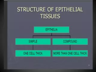
Index: Epithelial tissues Go directly to tissues Quick Quiz Epithelial types Simple squamous epithelium Simple Cuboidal epithelium Simple Columnar epithelium Stratified squamous epithelium Stratified cuboidal epithelium Pseudostratified columnar epithelium
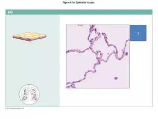
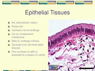
What type of epithelium is shown? The red arrows above point at a single layer of flattened cells that line a blood vessel. (The reddish stuff at the bottom of the screen is blood.) Note the flattened nuclei (dark ovals) which clue you in on the shape of the cells themselves.
Answer This is a simple squamous epithelium. There is another one at the orange arrow.
Answer This is also a simple squamous epithelium. This one is the epithelium that lines the renal corpuscles in the kidney.
FYI: This is a surface view of a simple squamous epithelium.
Here is an epithelium lining a duct. What type of epithelium is it?
Answer This is a simple cuboidalepithelium. The cells are about as tall as they are wide.
Answer This is a simple columnar epithelium. These light purple cells are clearly taller than they are wide.The dark purple ovals are their nuclei.
a The tissue indicated at “a” would be classified as…?
Answer This is a stratified squamous epithelium. This slide is a section of the tissue that lines your inner cheek.
a This is a slide of human skin. How would you classify the layer indicated by “a”?
Answer a This is also stratified squamous epithelium. If you look closely you will see multiple layers of flattened cells.
More info. The main difference between skin and the cells that line your cheek is that in skin, the outer layer of cells has become filled with a proteinacous substance called keratin. (become keratinized)
More info. The light purple layer of the epidermis looks very much like those cheek cells.
The epithelial layer which lines the cavity (white space) seen here would be classified as…?
…stratified cuboidal epithelium. Note that most of this cavity is lined by two layers of cells which are “block-like” in shape.
The epithelium at the surface (top) here would be classified as…
…pseudostratified columnar epithelium. Although the cells appear to be in several layers, they are actually all in contact with the basement membrane.
Quick Quiz For each of the following slides, try to guess which type of epithelium you are looking at. Before you check the answer, be able to justify to yourself why you think it is that type.
The outlined structure is a “tube” lined by Simple Cuboidal epithelium . There are also other similar tubes surrounding this one.
The epithelium which lines this “tube” would be classified as...
…stratified cuboidal epithelium.Notice that there are two layers of cells that are about as wide as they are tall.
The End Most of the images on this tutorial were taken from one of two excellent Histology sites on the Internet. Check them out if you want to see many more images of tissues. JayDoc HistoWeb: http://www.kumc.edu/instruction/medicine/anatomy/histoweb/ Loyola University Medical Education Network: Histology http://www.lumen.luc.edu/lumen/MedEd/Histo/frames/histo_frames.html I have links to both of these sites on my Web page under “Miscellaneous links”.