Download
1 / 49
560 likes | 1.22k Vues
Innate Immunity. Debra Laskin Professor and Chair Department of Pharmacology and Toxicology Ernest Mario School of Pharmacy laskin@eohsi.rutgers.edu. What is the Innate Immune Response?.

E N D
Innate Immunity Debra Laskin Professor and Chair Department of Pharmacology and Toxicology Ernest Mario School of Pharmacy laskin@eohsi.rutgers.edu
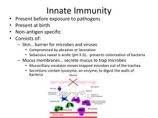
What is the Innate Immune Response? • Universal and evolutionarily conserved mechanism of host defense against infection; first line of defense; • Predates adaptive immune response • Found in all multicellular organisms (adaptive only in vertebrates) • Uses receptors and effectors that are ancient in their lineage • Provides protection against a wide variety of pathogens • Distinguishes self from non-self perfectly • Defects in innate immunity are very rare and almost always lethal
Elie Metchnikoff: “Father of Macrophages” 1908 Nobel prize Watched the reaction to a splinter inserted into a starfish. Hemocytes (amoeba-like cells) arrived and tried to ingest the foreign body; if they could not, they walled it off. Mammals do exactly the same thing to foreign bodies. Phagocytosis- to devour
Oyster hemocyte Mouse macrophage Similarity in appearance invertebrate and mammalian phagocyte striking; also use many of the same cytotoxic mechanisms (e.g., production ROS)
Innate Immunity: Functions • Provides a barrier to prevent the spread of infection • Physical • Skin (epithelial cells); Wounds, burns, insect bites • Mucosal surfaces (respiratory, GI, Reproductive) • Mechanical (tight junctions, movement) • Chemical (fatty acids, enzymes, pH, antimicrobial peptides) • Microbiological (normal flora)
Innate Immunity: Functions • Identifies and eliminates pathogens • Non-adaptive recognition systems • Activates molecules that target the microbe and aid in it’s identification • These factors may be surface expressed (TLR), released from immune cells (antibodies) or present within circulatory system (complement)
Innate Immunity: Functions • Initiates an inflammatory response • Reaction to injury or infection • Trauma to tissues or cells • Presence of foreign material (splinter) • Infectious agents (viruses, bacteria, fungi) • Delivers immune cells and effector molecules to the site of injury/infection • Components • Granulocytes, MP, inflammatory mediators • Blood vessels (endothelium) • Plasma proteins
Innate Immunity: Functions • Provides signals to alert the adaptive immune system to activate an effective specific immune response • Antigen processing and presentation (activation of T helper cells) • Upregulation of co-stimulatory molecules • MHC class II, CD80/86 • Induction of cytokine/chemokine response • IL-4, IL-12
Innate Immunity: Cellular Components • Granulocytes • Polymorphonuclear leukocytes (PMN, neutrophils) • Eosinophils • Basophils (blood) • Mast Cells (tissues) • Mononuclear Phagocytes (RES) • Monocytes (blood) • Macrophages (tissue)
Origin and Development of Macrophages and Granulocytes Bone Marrow Blood Tissue monoblast promonocyte monocyte macrophage basophilic promyelocyte basophil Phagocytic precursors eosinophilic promyelocyte mast cell eosinophil myeloblast neutrophilic promyelocyte Neutrophil (PMN)
Neutrophils(PMN) • Present in blood (60-70% of WBC) • Not normally present in tissues • Short lifespan - 12 hours • Functions: • First cell to respond to infection or injury (inflammatory site) • Ingest and kill pathogens • Release cytotoxic/proinflammatory mediators (ROS, proteolytic enzymes, bioactive lipids, chemokines)
MononuclearPhagocytes • Blood - monocytes (1-6% WBC) • Tissues - macrophages • mature form of monocytes • found in tissues (ex., gastrointestinal tract, lung, liver, brain, skin, spleen); reticuloendothelial system (RES) • Functions: • Inflammation- respond to injury, infection, other foreign substances • Phagocytize and kill pathogens • Wound repair, angiogenesis • Antigen presentation (activate adaptive immunity) • Tumor surveillance and cytotoxicity
Inflammatory Responses Injury or
Immediate release Initiate PMN rolling/adhesion Released 2 hours First Step: Activation of Vascular Endothelial Cells by Macrophages Endothelium • Form blood vessels • Macrophages present release TNFa and IL-1: upregulated expression of adhesion molecules on endothelium • P-selectin • E-selectin • ICAM-1 • Initiate PMN rolling, adherence (LFA-1) • Release chemokines (e.g., IL-8) induce PMN extravagation • PMN first to respond (within hours) • Monocyte second wave (24-48 hr)
Localization and Destruction of Pathogens and Foreign Substances Chemotaxis: migration to injured or infected site; mediated by chemotactic factors; complement, fMLP, C-X-C (PMN, IL-8) and C-C (mono, MCP-1) chemokines Phagocytosis: ingestion of foreign substances; receptor mediated, active process, requires energy
Localization and Removal of Foreign Substances Metabolic Destruction intracellular digestion, killing • Oxygen independent: defensins and granular cationic proteins, lactoferrin, lysozyme, acid hydrolases • Oxygen dependent: myeloperoxidase, hydrogen peroxide, superoxide anion, hydroxyl radicals, nitric oxide, peroxynitrite
Secretory Functions of Macrophages • Binding proteins (transferrin, fibronectin) • Complement components • Proteolytic enzymes (lysozyme) • Enzyme inhibitors (a2-macroglobulin) • Endogenous pyrogen (IL-1) • ROS (superoxide, hydrogen peroxide, hydroxyl radical) • RNS (nitric oxide, peroxynitrite) • Bioactive lipids (PAF, PG, LT, TBX) • Chemokines (C-C and C-X-C) • Growth factors (FGF, EGF, CSF) • Proinflammatory cytokines (IL-1, TNFa, IL-6) • Angiogenic factors: VEGF • Matrix remodeling proteins: TGFb, MMP
Stopping Inflammation: Wound Repair and Angiogenesis • Inflammatory macrophages release mediators that • Down regulate inflammation (eg., IL-10) • Inhibit inflammatory cell recruitment • Block specific immune responses • Initiate wound repair; matrix remodeling (MMPs, TGFb) • Recruit fibroblasts (FGF) • Induce angiogenesis (VEGF)
What Happens When Inflammation Fails to Resolve? • Frustrated Phagocytosis • Foreign Body Response • Macrophages wall off injurious agent • Chronic inflammation • Graunulomas • Tissue Injury • Cancer
Foreign Body Reactions Silicone droplets Suture material Clear globules of silicone released breast implant cannot be ingested; MP accumulate to wall off the material; note MP that has become a multi-nucleated giant cell showing the “asteroid bodies” characteristic of the foreign body reaction. Suture material is not digestible by macrophages, so it has been walled off by fibroblasts.
Bioactive Lipids ROI Activated Macrophage TNF-a IL-1 Proteolytic Enzymes RNI Macrophage Mediators Can Damage Host Tissues chemokines
H2O2 O2- OH- Lipid Peroxidation Membrane, Protein and DNA Damage Reactive Oxygen Species
Reactive Nitrogen Intermediates • Nitric oxide and peroxynitrite • Nitric oxide- formed from l-arginine by the enzyme nitric oxide synthase (NOS) • Macrophages: (NOSII) induced by inflammatory cytokines (IFNg, TNFa) and bacterially-derived products (LPS) • Highly labile; oxidizes nucleic acids, membranes, proteins • Nitric oxide reacts with superoxide anion forming peroxynitrite
Proinflammatory Cytokines • Tumor necrosis factor-a • Interleukin-1 • Interleukin-6 • Interleukin-18 • Chemokines • Interferon-g
Tumor Necrosis Factor-a • Proinflammatory • Primes phagocytes to produce ROI and RNI • Cytotoxic • Induces apoptosis and necrosis
Macrophage Processing of Antigens • Macrophages function as accessory cells or antigen processing cells (APC) • Macrophage associated antigen is 1000x more immunogenic • Processing of antigens involves change so that it binds MHC II (Ia) proteins; may involve unfolding, partial degradation, selection for epitope with high affinity for MHC II • Required for T-helper cell recognition of antigens • Other APC: B cells, epithelial cells, dendritic cells
How is it Possible for Macrophages to Perform all of these Functions? Macrophages functionally polarized into subpopulations by inflammatory signals in microenvironment M1 macrophages Activators: LPS, IFNg, TNFa, TLR ligands Cytotoxic (pathogens, tumor cells)/proinflammatory activity Release ROS, RNS, IL-12, TNFa, M1chemokines Promote Th1 responses M2 macrophages (alternatively activated) Activators: IL-4, IL-13, IL-10, immune complexes Antiinflammatory/wound repair activity Release IL-10, TGFb, PDGF, VEGF, MMP, EGF, FGF Immunosuppressive Promote Th2 responses
M2 Macrophages • M2a-activated by IL-4, IL13 • M2b- activated by immune complexes and TLR agonists or IL-1 • M2c (tumor associated macrophages)- activated by IL-10, TGFb, glucocorticoids **active in wound repair, angiogenesis, chronic inflammation
Tumor Associated Macrophages • Found within tumor microenvironment • M2 phenotype • Respond to cytokines (CSF-1) and chemokines (MCP-1) released by tumor cells • Hijacked by tumor cells to release mediators that contribute to tumor promotion, progression, angiogenesis and metastasis • EGF, VEGF, MMP, IL-1, IL-6, chemokines
How do Macrophages Identify Microbes? Pattern Recognition Receptors (PPR ) • Recognize pathogen associated molecular patterns (PAMP); conserved molecular patterns on microbes • Identify a class of microbes; ex., LPS, LTA, peptidoglycan, lipoarabinomannan, dsRNA, mannose, b-glycans • PAMP are often essential for microbe survival • Action Time • Immediate activation of effectors • Delays need for adaptive immunity
Pattern Recognition Receptors (PRR) Three broad classes based on expression profile, localization, function • PRR that signal an infection Toll Receptor Family • Expressed externally or internally • Binding activates “pro-inflammatory” signaling pathways • Phagocytic (endocytic) PRR • Expressed on the surface of phagocytic cells • Mediate uptake of microbe into phagocytes • Secreted PRR • Secreted by MP, epithelial cells, hepatocytes • Activate complement, opsonins, function as accessory proteins for PAMP recognition
Toll-like Receptor (TLR) Family First discovered in Drosophila Thirteen receptors identified in mice and humans Recognized motifs (PAMP) -lipopolysaccharide (LPS) from Gram-negative cell walls -peptidoglycans from the cell walls of both Gram-negative and Gram-positive bacteria -viral double-stranded RNA -CpG-rich bacterial DNA
Ligands are PAMP (pathogen- associated molecular patterns) Receptors are PRR (pattern- recognition receptors)
Single ligand-single response vs. multiple ligands-complex response
Accessory Proteins for TLR-4 Binding requires several accessory molecules • LBP/MD-2 (Macrophages) • RP105 / MD-1 (B cells) MD-2 MD-2 MD-2
Toll-like Receptor Signaling Resulting in the activation of gene transcription MD-2 IRAK MAPK
Intracellular PPR • Protein kinase receptor (PKR) • Activated upon binding to dsRNA (viruses) • Blocks viral and cellular protein synthesis (eIF2a) • Activates NF-kB, MAP kinase STAT & IRF signaling pathways • Induces apoptosis of infected cells and IFNa/b production • 2’-5’ Oligoadenylate Synthase and RNaseL • Family of IFN-inducible enzymes • Activated by dsRNA • RNaseL degrades viral and host RNA • Induces apoptosis
Intracellular PRR • Nucleotide-binding oligomerization domain (NOD) proteins -cytoplasmic surveillance proteins -bind peptidoglycans
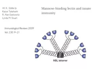
Surface Expressed PRRthat Bind Bacterial Carbohydrates • Mannose-binding receptor (C-type lectin) • Recognizes patterns of mannose residues in a certain spatial orientation unique to microbes • Only found on macrophages (not monocytes or PMN) • Glucan Receptor • Present on all phagocytes
Surface Expressed PRR thatBind other Bacterial Components • Scavenger Receptors • Recognize charged ligands • Polyanionic ligands (ds-RNA, LPS, LTA) • Acetylated low-density lipoproteins (LDL) • Found on all phagocytes (CD36; CD68) • MARCO (macrophage-specific, binds bacterial cell walls and LPS) • Phagocytosis of apoptotic cells • MFG-E8 (released from activated macrophages and binds to apoptotic cells via phosphatidylserine)
Secreted Pattern Recognition Molecules Important in complement activation Opsonization of microbial cells Primarily produced by the liver but can be produced by lung (SP) or phagocytes Acute Phase Proteins
Secreted Pattern Recognition Molecules • Collectins • Recognize microbial carbohydrate (CRD) domain Mannan-binding lectin Surfactant proteins (SP-A / SP-D) (lung) • Pentraxin • Recognize phosphorylcholines on microbes; • Lipid Transferases • Recognizes peptidoglycans • Peptidoglycan recognition proteins (PGRS) • LPS binding protein (LBP
INNATE IMMUNITY PHYSICAL BARRIERS Skin, mucous membrane CHEMICAL BARRIERSpH, lipids, enzymes CELLSgranulocytes, monocytes, macrophages ADAPTIVE IMMUNITY HUMORAL B cells antibodies CELL MEDIATED T cells lymphokines MP
Cellular Interactions in the Immune System Activated MP MP (+) PA Sensitized Antigen destruction Th1 (+) Th Th MP (+) AG (-) Y Tcyt Th2 (+) Treg (-) (-) B plasma Sensitized B Bmem