Download
1 / 1
10 likes | 153 Vues
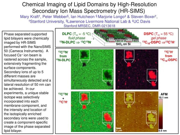
Chemical Imaging of Lipid Domains by High-Resolution Secondary Ion Mass Spectrometry (HR-SIMS) Mary Kraft † , Peter Webber § , Ian Hutcheon § Marjorie Longo ‡ & Steven Boxer † , † Stanford University, § Lawrence Livermore National Lab & ‡ UC Davis Stanford MRSEC, DMR-0213618.

E N D
Chemical Imaging of Lipid Domains by High-Resolution Secondary Ion Mass Spectrometry (HR-SIMS) Mary Kraft†, Peter Webber§, Ian Hutcheon § Marjorie Longo‡& Steven Boxer†, †Stanford University, §Lawrence Livermore National Lab & ‡UC Davis Stanford MRSEC, DMR-0213618 SiO2 on Si DLPC (Tm = -5 ºC) fluid phase 15N-DLPC 12C15N- DSPC (Tm = 55 ºC) gel phase 13C18-DSPC 13C1H- Phase separated supported lipid bilayers were chemically imaged by HR-SIMS performed with the NanoSIMS 50 (Cameca Instruments). A focused Cs+ ion beam is rastered across the sample, extensively fragmenting the surface components. Secondary ions of up to 5 different masses are simultaneously detected and a lateral resolution of 50 nm can be achieved. In our experiments, a unique stable isotope was selectively incorporated into each membrane component, and the intensity and location of the isotopically enriched secondary ions were used to create a component-specific image of the phase separated lipid bilayer. 13C1H- from 13C18-DSPC 12C15N- from 15N-DLPC 12C15N- + 13C1H- AFM