Download
1 / 76
980 likes | 1.98k Vues
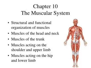
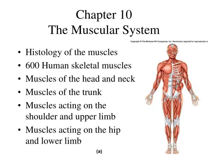
Chapter 10 The Muscular System. Histology of the muscles 600 Human skeletal muscles Muscles of the head and neck Muscles of the trunk Muscles acting on the shoulder and upper limb Muscles acting on the hip and lower limb. Introduction to Muscle.

E N D
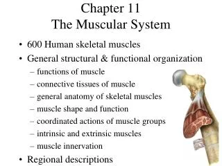
Chapter 10The Muscular System • Histology of the muscles • 600 Human skeletal muscles • Muscles of the head and neck • Muscles of the trunk • Muscles acting on the shoulder and upper limb • Muscles acting on the hip and lower limb
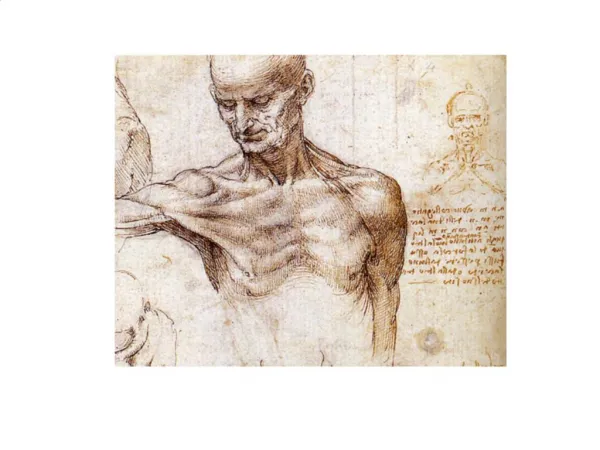
Introduction to Muscle • Movement is a fundamental characteristics of all living things • Cells capable of shortening & converting the chemical energy of ATP into mechanical energy • Types of muscle • skeletal • cardiac • smooth • Physiology of skeletal muscle • basis of warm-up, strength, endurance & fatigue
Muscle Fibers (Form follows Function) • Multiple flattened nuclei against inside of plasma membrane • due to fusion of multiple myoblasts during development • unfused satellite cells nearby can multiply to produce a small number of new myofibers • Sarcolemma has tunnel-like infoldings or transverse (T) tubules that penetrate the cell • carry electric current to cell interior • Sarcoplasm is filled with • myofibrils (bundles of parallel protein microfilaments called myofilaments) • glycogen for stored energy & myoglobin binding oxygen • Sarcoplasmic reticulum is series of interconnected, dilated, calcium storage sacs called terminal cisternae
Thick Filaments • Made of 200 to 500 myosin molecules • 2 entwined polypeptides (golf clubs) • Arranged in a bundle with heads (cross bridges) directed outward in a spiral array around the bundled tails • central area is a bare zone with no heads
Thin Filaments • Two intertwined strands of fibrous (F) actin • each subunit is a globular (G) actin with an active site • Groove holds tropomyosin molecules, each blocking the active sites of 6 or 7 G actins • One small, calcium-binding troponin molecule stuck to each tropomyosin molecule
Elastic Filaments • Huge springy protein called titin (connectin) • runs through core of each thick filament • connects thick filament to Z disc structure • Functions • keep thick & thin filaments aligned with each other • resist overstretching • help the cell recoil to its resting length (elasticity)
Regulatory & Contractile Proteins • Myosin & actin are contractile proteins (they do work) • Tropomyosin & troponin are regulatory proteins • act like a switch that starts & stops shortening of muscle cell • the release of calcium into sarcoplasm and its binding to troponin, activates contraction • troponin moves the tropomyosin off the actin active sites
I A I Striations = Organization of Filaments • Dark A bands (regions) alternating with lighter I bands (regions) • anisotrophic (A) and isotropic (I) stand for the way these regions affect polarized light • A band is thick filament region • lighter, central H band area contains no thin filaments • I band is thin filament region • bisected by Z disc protein called connectin, anchoring elastic & thin filaments • from one Z disc (Z line) to the next is a sarcomere
Relaxed versus Contracted Sarcomere • Muscle cells shorten because their individual sarcomeres shorten • pulling Z discs closer together • pulls on sarcolemma • Notice neither thick nor thick filaments change length during shortening • Their overlap changes as sarcomeres shorten
Skeletal muscle • striations & peripheral nuclei
Smooth muscle • lack of striations & central nuclei
Cardiac muscle • striations, intercalated discs & central nuclei
Parts of a Skeletal Muscle • Origin • attachment to stationary end of muscle • Belly • thicker, middle region of muscle • Insertion • attachment to mobile end of muscle
Skeletal Muscle Shapes • Fusiform muscles • thick in middle & tapered at ends • biceps brachii m. • Convergent muscle • broad at origin and tapering to a narrower insertion • Parallel muscles • parallel fascicles • rectus abdominis m.
Skeletal Muscle Shapes (2) • Circular muscles • act as sphincters • ring around body opening • orbicularis oris • Pennate muscles • fascicles insert obliquely on a tendon • unipennate, bipennate or multipennate • palmar interosseus, rectus femoris & deltoid
Coordinated Muscle Actions Example • Prime mover or agonist • produces most of force • Synergist aids the prime mover • stabilizes the nearby joint • modifies the direction of movement that occurs • Antagonist • opposes the prime mover • preventing excessive movement and injury • Fixator • prevents movement of bone that prime mover is attached to
Muscle Actions during Elbow Flexion • Prime mover (agonist) = biceps brachii m. • Synergist = brachialis m. • Antagonist = triceps brachii m. • Fixator = muscle that holds scapula firmly in place such as rhomboideus m. Definitions
Intrinsic and Extrinsic Muscles • Intrinsic muscles are contained within a region such as the hand. • Extrinsic muscles move the fingers but are found outside the region.
How Muscles are Named • Nomina Anatomica • system of Latin names developed in 1895 • updated since then • English names for muscles are slight modifications of the Latin names. • Table 10.1 = terms used to name muscles digiti = of a finger levator = elevates a body part profundus = deepest quadriceps = having 4 heads
Learning Strategy • Explore the location, origin, insertion and innervation of 160 skeletal muscles using the tabular information in this chapter. • Increase your retention & understanding by: • examining models and photographic atlases • palpating yourself using the images in Atlas B • observe an articulated skeleton • say the names aloud and check your pronunciation
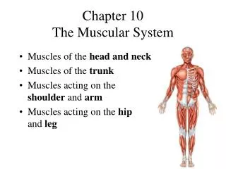
Muscles of Facial Expression • Small muscles that insert into the dermis • Innervated by facial nerve (CN VII) • Paralysis causes face to sag • Found in scalp, forehead, around the eyes, nose and mouth, and in the neck
Muscles of the Scalp and Forehead Frontalis Occipitalis Occipitofrontalis is found in the scalp. Frontalis m. raises the eyebrows while Occipitalis m. fixes the galea aponeurotica
Muscles around the Eyes Corrugator supercilii Procerus Orbicularis Oculi Nasalis Orbicularis oculi closes the lips. Corrugator draws the eyebrows together. Procerus pulls down the skin of forehead. Nasalis widens nostrils.
Muscles around the Mouth • Orbicularis oris encircles mouth & other mm blend into it • Levator & depressor of labii (lip) & anguli (angle of mouth) • Risorius & zygomaticus curl corner of mouth up in smile • Buccinator keeps food on top of teeth, blowing & sucking Levator labii superioris Zygomaticus major Buccinator Risorius Depressor anguli oris Orbicularis oris Depressor labii inferioris
Muscles of Mastication • 4 Major muscles • Arise from skull & insert on mandible • Temporalis & Masseter elevate the mandible • Medial & Lateral Pterygoids help elevate, but produce lateral Swinging of jaw used to grind with molars Temporalis Masseter Lateral pterygoid Medial pterygoid
Muscles of Respiration • Breathing requires the use of muscles • diaphragm • external intercostal muscles • internal intercostal muscles • Contraction of the first 2 produces Inspiration • Contraction of the last produces Forced Expiration • Normal Expiration requires little muscular activity • elastic recoil of tissues and gravity collapsing the chest • only inspiratory muscles active in braking action, so exhalation is smooth
Muscles of Respiration -- Diaphragm Central tendon • Muscular dome between thoracic and abdominal cavities • Muscle fascicles extend to a fibrous central tendon • Contraction flattens it • increases the vertical dimension of the thorax drawing air into the lungs • raises the abdominal pressure to help expel urine, feces and facilitating childbirth
Muscles of Respiration -- Intercostals • External intercostals • extend downward and anteriorly from rib to rib • pull ribcage up & outward during inspiration • Internal intercostals • extend upward and anteriorly from rib to rib • pull ribcage downward during forced expiration
Muscles of Respiration - Serratus • Serratus posterior superior • elevates ribs 2-5 during inspiration • Serratus posteriori inferior • depresses ribs 9-12 during inspiration
Muscles of the Abdomen • 4 Pairs of sheetlike muscles • external oblique • internal oblique • transverse abdominis • rectus abdominis • Functions • support the viscera • stabilize the vertebral column • help in respiration, urination, defecation & childbirth
External oblique superficial downward anteriorly inguinal ligament Rectus abdominis vertical, straplike tendinous intersections rectus sheath linea alba Rectus Abdominis & External Oblique Rectus abdominis External oblique
Internal oblique anteriorly upwards Transverse abdominis horizontal fiber orientation deepest layer Internal Oblique -Transverse Abdominis Transverse abdominis Internal oblique
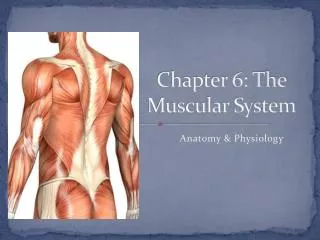
Superficial Muscles of the Back Semispinalis Splenius Infraspinatus Levator scapulaeRhomboideus Supraspinatus Teres major Gluteus maximus Gluteus medius Trapezius Latissimus dorsi
Muscles Acting on the Pectoral Girdle • Originate on axial skeleton & insert onto clavicle or scapula • Anterior muscle group = 2 muscles • Posterior muscle group = 4 muscles • Scapular movements produced include • medial and lateral rotation of the scapula • elevation and depression of the scapula • protraction and retraction of the scapula • Clavicle braces the shoulder & limits movement
Pectoralis Minor ribs 3-5 to coracoid process of scapula protracts & depresses scapula lifts ribs during forced expiration Serratus Anterior ribs 1-9 to medial border of scapula abducts & rotates or depresses scapula throwing muscle Anterior Scapular Muscle Group