Download
1 / 39
490 likes | 1.35k Vues
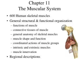
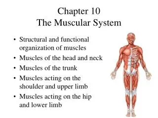
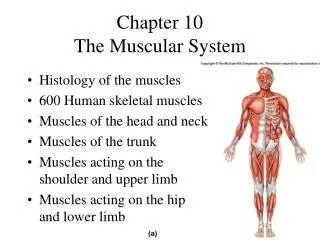
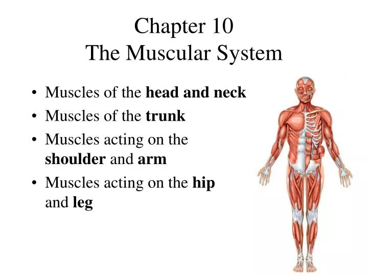
Chapter 10 The Muscular System. Muscles of the head and neck Muscles of the trunk Muscles acting on the shoulder and arm Muscles acting on the hip and leg. The Functions of Muscles. 600 Human skeletal muscles Movement of body parts and organ contents

E N D
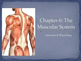
Chapter 10The Muscular System • Muscles of the head and neck • Muscles of the trunk • Muscles acting on the shoulder and arm • Muscles acting on the hip and leg
The Functions of Muscles • 600 Human skeletal muscles • Movement of body parts and organ contents • Maintain posture and prevent movement • Communication - speech, expression & writing • Control of openings and passageways • Body heat production
Connective Tissues of a Muscle Tendon Deep fascia Epimysium Perimysium Endomysium
Connective Tissues of a Muscle • Epimysium • covers whole muscle belly • Perimysium • surrounds a bundle of cells called a fascicle • Endomysium • thin layer surrounding each cell • oPEN
Muscle Attachments • Direct attachment to bone • epimysium is continuous with periosteum • intercostal muscles • Indirect attachment to bone • epimysium continues as tendon • biceps brachii
Parts of a Skeletal Muscle • Origin • attachment to stationary end of muscle • First in a muscle name (if the name allows it) • Insertion • attachment to mobile end of muscle • End of the muscle name (if the name allows it)
Coordinated Muscle Actions • AGONIST: the muscle that causes a desired action, also called the Prime Mover. • ANTAGONIST: produces an opposite action (Anti to agonist) • SYNERGIST: steady a movement, thus preventing unwanted movements and helping the agonist • the extensor carpi muscles act as synergists for the flexor digitorum muscles (Try it) • FIXATORS: synergist muscles which stabilize the origin of the agonist so that it can act more efficiently (shoulder muscles)
Muscle Actions during Elbow Flexion • Prime mover (agonist) = biceps brachii m. • Synergist = brachialis m. • Antagonist = triceps brachii m. • Fixator = muscle that holds scapula firmly in place such as rhomboideus m. * *
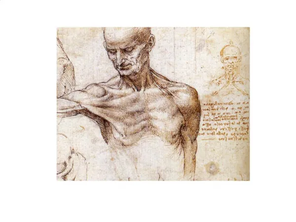
Learning Strategy • Know the names of the muscles, say the names aloud (check your pronunciation) while pointing to them on yourself • Know the Origin/ Insertion and Action • Using the information in this chapter. • Increase your retention & understanding by: • examining models and photographic atlases • palpating yourself using the images in Atlas B • observe an articulated skeleton
Muscles of the Face I(Fig. 10.7) Frontalis Occipitalis Occipitofrontalis is found in the scalp. Frontalis m. raises the eyebrows while Occipitalis m. fixes the galea aponeurotica
Muscles of the Face II(Fig. 10.7) • Orbicularis oris encircles mouth (Orbicularis = round circle) • Masseter chews • Orbicularis oculi closes the eye. Masseter Orbicularis oris
Q. What Muscle?(Think of your Aunt’s Sunday Dinner) • A. The platysma muscle makes us grimace
Shoulder muscles I(Fig. 10.17) Upper Trapezius Deltoid Middle and lower Trapezius
Shoulder Muscle II(Fig. 10.15 a) Pectoralis major
Pectoralis Minor ribs 3-5 to coracoid process of scapula Deep to ________ Serratus Anterior ribs 1-8 to medial border of scapula Shoulder muscles III (Fig. 10.15 b)
SITS muscles (Rotator Cuff) Supraspinatus- Infraspinatus Teres minor Subscapularis SITS Muscles(Fig 10.22) Supraspinatus Subscapularis “M” medial rotation of humerus Infraspinatus Teres minor “L” lateral rotation of humerus
ARM MUSCLES(Fig 10.22) • Principal flexor • biceps brachii • inserts on radius and ulna • Two origins and insertions • Synergistic flexor • brachioradialis • Teres Major • Majors are below the minors • Prime extensor • triceps brachii • inserts onto ulna • Three origins
Digitorum = inserts into fingers Carpi = inserts onto metacarpal bones Pollicis (means thumb)= inserts into thumb Intrinsic is of the hand, extrinsic is in the forearm Brevis = short, Longus = ________ Ulnaris = on ulna side Radialis = on ________ side Thenar group = fleshy base of thumb muscles Hypothenar group = base of little finger muscles Hand/ wrist Extensor and FlexorsTerms
Rhomboideus mm. Latissimus Dorsi Back Muscles(Fig. 10.17)
HIP MUSCLES I(Fig. 10.34) Gluteus medius • Gluteus maximus • forms mass of the buttock • prime hip extensor • provides most of lift when you climb stairs Gluteus maximus
Gluteus minimus HIP MUSCLES II(Fig. 10.31) Piriformis • Deep muscles of the hip • Important in walking to shift body weight when foot is lifted • Sciatic nerve lines deep to the ______ muscle
HIP MUSCLES III (Fig. 10.30)Iliopsoas • Iliopsoas muscle • crosses anterior surface of hip joint & inserts on femur • iliacus portion arises from iliac fossa • psoas portion arises from lumbar vertebrae (L1-L5) • major hip flexor Iliopsoas
HIP MUSCLES IV(Fig 10.30) Adductors of the Hip Joint • Adductor magnus is also an extensor of hip joint • Pectineus, Adductor brevis and Adductor longus adduct the femur • (Just know them generally as adductors) Pectineus Adductor brevis Adductor longus Adductor magnus
LEG MUSCLES I(Fig. 10-32 b) • QUADS • 4 headed muscle attaches to tibial tuberosity • extends knee joint • rectus femoris arises from ilium so flexes hip joint • 2 joint muscle • quadriceps femoris tendon attaches to patella • patellar ligament attaches to tibia
LOWER LEG MUSCLES I(Fig. 10.35) Tibialis anterior • ANTERIOR • Extensor hallucis longus = extension of big toe & ankle • Tibialis anterior = dorsiflexes and inverts foot Extensor hallucis longus
LOWER LEG MUSCLES II(Fig. 10.37) Gastrocnemius Soleus • POSTERIOR • Gastrocnemius = flexes knee and plantar flexes ankle • Soleus = plantar flexes ankle
Diaphragm (Fig. 10.13)Rectus Abdominis(Fig. 10.15) • Rectus abdominis • vertical, straplike • tendinous intersections • LINEA ALBA • Diaphragm • Muscular dome between thoracic and abdominal cavities, breathing
A Diamond and Two Triangles • Muscles of the Pelvic Floor are in a region called the perineum or pelvic diaphragm • One diamond and two triangles • diamond region bounded by pubic symphysis, coccyx and ischial tuberosities • urogenital triangle • anterior • contains the urethra & vagina • anal triangle • posterior • contains the anal canal
Pelvic diaphragm = 2 muscles levator ani m. supports viscera & functions during defecation Muscle of the Pelvic Diaphragm Levator ani
General Muscles of the Deep Back Erector spinae group
Athletic Injuries • Proper conditioning and warm-up is needed • Common injuries • shinsplints • pulled hamstrings • tennis elbow • Treat initially with rest, ice, compression and elevation (RICE) • “No pain, no gain” is a dangerous misconception.
Hernias • Protrusion of viscera through muscular wall of abdominopelvic cavity • Inguinal hernia • most common type of hernia (rare in women) • viscera enter inguinal canal or even the scrotum • Umbilical hernia • viscera protrude through the navel
Surface Anatomy Anatomy Game Show