Download
1 / 36
420 likes | 756 Vues
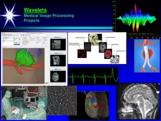
Corso di Modellazione e Simulazione di Sistemi Fisiologici. Medical Image Processing. Federica Caselli. Department of Civil Engineering University of Rome Tor Vergata. Medical Imaging. X-Ray. Ultrasound. CT. PET/SPECT. MRI. Digital Imaging!. Medical Image Processing. What kind ?.

E N D
CorsodiModellazione e SimulazionediSistemiFisiologici Medical Image Processing Federica Caselli Department of Civil Engineering University of Rome Tor Vergata
Medical Imaging X-Ray Ultrasound CT PET/SPECT MRI Digital Imaging!
Medical Image Processing Whatkind? Whatfor? • Image compression • Image denoising • Image enhancement • Image segmentation • Image registration • Image fusion • Image storage, retrieval, transmission • Telemedicine • Quantitative analysis • Computer aided diagnosis, surgery, treatment and follow up Tonamebut a few! Image analysis software are becoming an essential component of the medical instrumentation
Two examples Mammographicimagesenhancement and denoisingforbreastcancerdiagnosis Delineationof target volume forradiotheraphy in SPECT/PETimages
Mammographic image enhancement Shape Boundary Diseasesigns in mammograms: MASSES EARLY DIAGNOSIS IS CRUCIAL FOR IMPROVING PROGNOSIS!
Mammographic image enhancement In 60-80 %ofbreastcancers at hystologicalexamination Diseasesigns in mammograms: MICROCALCIFICATIONS Morphology, size (0.1 - 1 mm), number and clusters INTERPRETING MAMMOGRAMS IS AN EXTREMELY COMPLEX TASK EARLY DIAGNOSIS IS CRUCIAL FOR IMPROVING PROGNOSIS!
Transformed-domain processing Image Enhancedimage Transformed domain representation Modifiedimage in transformed domain 1) 2) 3) T T-1 E(x) Transform Transformed-domain processing Inverse Transform Transformed-domain processing: signal is processed in a “suitable” domain. “Suitable” depends on the application
Fourier-based processing S + N S: 200 Hz N: 5000 Hz |H(ω)| |Y(ω)| |X(ω)| LPF Is it suitable for mammographic image processing?
Fourier-based processing Fourier is extremely powerful for stationary signals but No time (or space) localization ?
Short-Time Fourier Transform Frequency and time domain information! However a compromise is necessary...
Short-Time Fourier Transform Frequency Time Narrow window
Short-Time Fourier Transform Frequency Time Medium window
Short-Time Fourier Transform Frequency Wavelet Transform: more windows, with suitable time and frequency resolution! Time Once chosen the window, time and frequency resolution are fixed Large window
Wavelet Transform u s “If you painted a picture with a sky, clouds, trees, and flowers, you would use a different size brush depending on the size of the features. Wavelet are like those brushes.” I. Daubechies
Wavelet Transform “If you painted a picture with a sky, clouds, trees, and flowers, you would use a different size brush depending on the size of the features. Wavelet are like those brushes.” I. Daubechies
Wavelet Transform “If you painted a picture with a sky, clouds, trees, and flowers, you would use a different size brush depending on the size of the features. Wavelet are like those brushes.” I. Daubechies
Wavelet Transform “If you painted a picture with a sky, clouds, trees, and flowers, you would use a different size brush depending on the size of the features. Wavelet are like those brushes.” I. Daubechies
Wavelet Transform “If you painted a picture with a sky, clouds, trees, and flowers, you would use a different size brush depending on the size of the features. Wavelet are like those brushes.” I. Daubechies
Wavelet Transform “If you painted a picture with a sky, clouds, trees, and flowers, you would use a different size brush depending on the size of the features. Wavelet are like those brushes.” I. Daubechies Many type of Wavelet Transform (WT): Continuous WT and Discrete WT, each with several choices for the mother wavelet. Moreover, Discrete-Time Wavelet Transform are needed for discrete signals
Dyadic Wavelet Transform S. Mallat and S. Zhong, “Characterizationofsignalsfrommultiscaleedge”, IEEE Transactions on Pattern Analysis and Machine Intelligence, Vol. 14, No. 7, 1992.
Implementation Algorithme à trous Higherscales d1 G() d2 ao G(2) d3 H() G(4) H(2) a1 H(4) a2 a3 Decomposition Discrete-timetransform
Implementation Algorithme à trous K() ao K(2) H() K(4) H(2) a1 H(4) a2 Reconstruction Discrete-timetransform Higherscales d1 G() d2 ao G(2) d3 H() G(4) H(2) a1 H(4) a2 a3 Decomposition
Filters r = 1 GGradientfilter
Filters r = 2 GLaplacianfilter
1D Transform Scale Signal GRADIENTE LAPLACIANO Detailcoefficients
Denoising Segnale rumoroso outlier W Segnale ricostruito W-1
Wavelet Thresholding Hard thresholding Soft thresholding Key issue: thresholdsselection
Implementation G(x) K(x) L(y) do1 ao ao G(y) L(x) K(y) dv2 G(2x) K(2x) L(2y) do2 H(x) H(y) H(x) H(y) G(2y) L(2x) K(2y) a1 a1 H(2x) H(2y) H(2x) H(2y) a2 Decomposition Reconstruction Algorithme à trous Discrete-timetransform dv1
DDSM 5491 x 2761 12 bpp Resolution: 43.5 m 4.45 cm ROI 1024 x 1024 * University of South Florida, http://marathon.csee.usf.edu/Mammography/Database.html
Masses dv do m Scale 1 2 3 4
Microcalcifications dv do m 1 2 3 4
Wavelet-based signal processing Image Enhancedimage Wavelet coefficients Modifiedcoefficients 1) 2) 3) W E(x) W-1 Decomposition Enhancement Reconstruction Enhancingverticalfeatures Varying the gain G=8 G=20 Extremelysimple and powerfultoolforsignalprosessing. Manymanyapplications! Linear enhancement
Wavelet-based signal processing Mammogramshave low contrast E(x) Mustbeadaptive and automatic G T1 T2 Riskregion Saturationregion Amplificationregion Key issue: operator and thresholdsselection