Download
1 / 19
1.65k likes | 3.74k Vues
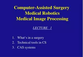
CVIP Lab. The University of Louisville. Medical Image Processing. M. Sabry Hassouna Ph.D. Computer Vision & Image Processing Laboratory (CVIP) Louisville, Kentucky. Medical Imaging. The study of medical imaging is concerned with

E N D
CVIP Lab The University of Louisville Medical Image Processing M. Sabry Hassouna Ph.D. Computer Vision & Image Processing Laboratory (CVIP) Louisville, Kentucky
Medical Imaging The study of medical imagingis concerned with • Interaction of all forms of radiation with tissue. • The development of appropriate technology to extract clinically useful information, usually in the form of an image from the observed technology.
X-Ray • Image Formation • A beam of X-rays is directed through a patient onto a film. • The film provides a measure of the ray attenuation in tissue. Nov 8, 1895 Wilhelm Konrad Röntgen reported discovery of new “rays” (Nobel Prize in physics in 1901). Jan 13, 1896 First clinical use of X-rays by 2 British doctors to find a needle in a hand. + Excellent for imaging bones. - No depth information, bad for soft tissue, excessive radiation
Computed Tomography (CT) Image Formation The object is viewed from a number of different angles and then a cross-sectional image of it can be computed (reconstructed). Nov 8, 1895 G. Hounsfield (computer expert) and A.M Cormack (physicist) (Nobel Prize in Medicine in 1979). + Provide 3D anatomical information + Preserves topology (bones) - Excessive radiation - Not good for all soft tissues
slice-by-slice scanning Spiral (volume) scanning (Very Fast) CT Acquisition Techniques 3D Reconstruction
Magnetic Resonance Imaging (MRI) Image Formation - Hydrogen nuclei (protons) under a strong magnetic field spin in phase with one another and align with the field. - Relaxed protons induce a measurable radio signal. 1952 F. Bloch and E. Purcel, extended by R. Ernst) (Bloch & Purcel: Nobel Prize in Physics in 1952) (Ernst: Nobel Prize in Chemistry in 1991) + Main modality for image guided surgery. + Superb ability to discriminate between subtle differences in tissue characteristics. + Very safe. - Less accurate for bone scanning.
Ultrasound (US) Image Formation An ultrasonic energy is propagated into the patient from a transducer placed on the skin and back-scattered echo signal is recorded by the same transducer. 1979: Samuel H. Maslak + Noninvasive + Clean & safe + In-expensive - Noisy - Gas filled and bony structures cannot be imaged because they absorb ultrasound waves.
Positron Emission Tomography (PET) Image Formation - Detection of radiation from the emission of positrons. 1998-2001: Dr. David Townsend and Dr. Ron Nutt. + Valuable technique for some diseases and disorders. + Amount of radiation is small - Invasive (inject radioactive material)
Magnetic Resonance Angiography (MRA) Image Formation Imaging the blood vessels (moving spins) using MRI scanner. TOF (time-of-flight) PCA (phase contract angiography) (CEA) (contrast enhanced angiography) (CTA) (computer tomography angiography) (DSA) (digital subtraction angiography) DSA CTA TOF
Data Processing 1. Preprocessing Filtering, registration 2. Detection Finding objects (nodules, polyps, organs) 3. Segmentation Exact delimitation of objects 4. Analysis Measurement (volume, curvature) 5. Classification/diagnoses
Preprocessing Original Enhanced
Detection Find location of objects of interest without prior knowledge about their location/existence • Bones • Organs • Polyps in colon • Nodules in lungs
Segmentation Exactly delimitate objects, once they are detected. - Vessels - Liver - Cardiac imaging (left ventricle) - Brain
Analysis • Measurement Volume - growth Vessel stenosis • Functional imaging Stroke Cardiac perfusion Tumor perfusion • Cardiac function LV motion Injection fraction
Classification / Diagnoses • Comparison to developed atlases • Use of knowledge databases • Classify as normal/abnormal (brain) • Classify lung nodules as benign/malignant • Determine cancer/non-cancer