Download
1 / 20
210 likes | 340 Vues
GENERAL STRUCTURE OF DIGESTIVE TRACT. Functional layers of GIT. The gastrointestinal tract has four distinct functional layers: mucosa , submucosa , muscularis propria adventitia . . Mucosa. Innermost coat. epithelium , lines the luminal surface

E N D
Functional layers of GIT • The gastrointestinal tract has four distinct functional layers: • mucosa, • submucosa, • muscularispropria • adventitia.
Mucosa.Innermost coat. • epithelium, lines the luminal surface • lamina propria (supporting)- mucosal glands, B/V, lymph vessels, mucosa assosiated lymphoid tissue (MALT), macrophages,eosinophils, fenestrated capillaries. • muscularismucosae, (smooth muscle layer)which produces local movement and folding of the mucosa. • At four points along the tract, the mucosa undergoes abrupt transition from one form to another: the gastro-oesophageal junction, the gastroduodenal junction, the ileocaecal junction and the recto-anal junction.
Submucosa. Between mucosa and muscular externa • This layer of loose collagenous supporting tissue supports the mucosa and contains the larger blood vessels, lymphatics and nerve plexus (submucosal nerve plexus -Meissners plexus). • No glands are present in submucosa except in stomach and duodenum.
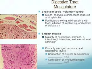
Muscularispropria. The muscular wall proper consists of smooth muscle that is usually arranged as an • inner circular layer (tight spiral)-constricts the gut lumen. • an outer longitudinal layer (loose spiral)-shorten the gut (widens the lumen). • In the stomach only, there is an inner oblique layer of muscle. The action of the two layers, at right angles to one another, is the basis of peristaltic contraction. • Nerve plexus (Myenteric plexus also called as Auerbach’s plexus)
Adventitia. • Rich in elastic fibres. • This outer layer of loose supporting tissue conducts the major vessels, nerves and variable adipose tissue. • Where the gut lies within the abdominal cavity (peritoneal cavity), the adventitia is referred to as the serosa (visceral peritoneum) and is lined by a simple squamous epithelium (mesothelium). • Elsewhere, the adventitial layer merges with retroperitoneal tissues.
Four mucosal types Secretory. This type occurs only in the stomach. The mucosa consists of long, closely packed tubular glands that are simple or branched depending on the region of the stomach. Absorptive. This mucosal form is typical of the entire small intestine. The mucosa is arranged into finger-like projections, called villi, which increase surface area with intervening short glands called crypts. In the duodenum, some crypts extend through the muscularismucosae to form submucosal glands called Brunner's glands. This is the major histological feature that differentiates the duodenum from the jejunum and ileum. Absorptive/protective. This form lines the entire large intestine. The mucosa is arranged into closely packed, straight tubular glands consisting of cells specialised for water absorption and mucus-secreting goblet cells to lubricate the passage of faeces.