Download
1 / 20
200 likes | 325 Vues
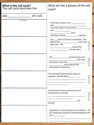
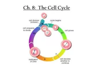
Cell Cycle – “The Hourly Life of a Cell” What happens when and how. Why do cells divide? To make a new organism Growth Repair Replacement of normal cell loss Development. The Cell Cycle. Goes back to normal functions (making proteins, etc. 2 new cells form. Hours to days

E N D
Cell Cycle – “The Hourly Life of a Cell”What happens when and how • Why do cells divide? To make a new organism Growth Repair Replacement of normal cell loss Development
The Cell Cycle Goes back to normal functions (making proteins, etc. 2 new cells form Hours to days Making proteins Normal cell functions Growth 2 Growth 1 G0 Makes an exact copy of DNA 3-6 hours
Apoptosis – Programmed Cell Death • Nucleases and Proteases are specifically activated • Time activated apoptosis important for normal development. Dev. of nervous system Dev. of immune system Hand and food dev. Leaf termination
Chromatin vs. Chromosomes • Chromatin – 2 m of DNA must fit in a 1x10⁻⁵ m nucleus. DNA wrapped around histone proteins to organize it and allow it fit into the nucleus • Remember – it is condensed 200,000 x to fit in the nucleus • It is still loosely coiled enough that enzymes can get into the DNA to copy it and make mRNA for protein synthesis • It is the normal form of DNA during all phases of the cell cycle except mitosis
Chromosomes • DNA compacted 12,000 times from chromatin • Cannot read or copy the DNA in chromosomes – it is too tightly wound • Formed solely during mitosis in order to divide the doubled DNA in ½
Formation of Chromatin and Chromosomes Chromatin Up Close
Structure of the Mitotic Chromosome Showing Sister Chromatids, Centromeres, Kinetochores, and Spindle Fiber Attachment Chromatid – ½ of a chromosome Sister chromatid – each half of the same chromosome Centromere – complex of proteins attached to DNA holding the sister chromatids together Kinetochore – complex of proteins attached to the outside surface of the chromosome at the centromeric region – where spindle fibers attach
Interphase Stages of Mitosis • Interphase is not part of mitosis – it is the time between cell divisions • Interphase includes G1, S, and G2 • During interphase the cell is doing its normal metabolic activities like protein synthesis • The cells are performing their duty as part of a tissue • The DNA duplicates to get ready for mitosis • The DNA is in chromatin form
Prophase • The chromatin begins to condense into chromosomes and become visible in the nucleus • The nuclear membrane begins to break down • Centrosomes duplicate, form spindles, & move to the poles • Proteins attach to chromosomes forming kinetochores • Spindle fibers attach to the kinetochores and chromosomes begin moving
Metaphase The chromosomes are lined up down the equator by the spindles
Anaphase • The sister chromatids separate at the centomeres • Each chromatid (now called a chromosome) heads to the pole of the cell • The movement is due to kinetochore movement along the spindle fiber microtubules
Telophase • The chromosomes are completely to the opposite poles • New membranes start to form around the DNA • The chromosomes begin to decondense back to chromatin • Cytoplasm begins to pinch in animal cells and a cell wall begins to form in plant cells – This is cytokinesis http://www.bioweb.uncc.edu/biol1110/Stages.htm
Interphase After telophase is complete, the cells reenter interphase and go about their normal business The DNA is totally decondensed, new nuclei reformed, and there are totally 2 new cells
Mitosis Quiz – Animal Cells Interphase Metaphase Anaphase Interphase Prophase Telophase
Mitosis Quiz – Plant Cells Metaphase Telophase Anaphase Interphase Prophase Interphase http://biology.nebrwesleyan.edu/benham/mitosis/
Differences Plant vs. Animal Cell Mitosis • Plant cells do not have centrioles in their centrosomes but animal cells do ????? • Plant cells cannot pinch in due to the cell wall – a new cell wall forms down the middle from the endoplasmic ret. • Plant cells divide slower due to having to reform the cell wall
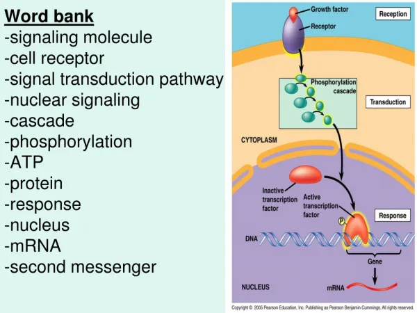
Control of The Cell CycleRegulated by internal and external signals Internal Signals • There are checkpoints at the end of G1 and end of G2. Signal molecules cause the cycle to go on or stop. • Signal examples Protein kinases (static levels) + cyclins (conc. fluctuates) = active kinases MPF is an activated kinase that promotes G2→M Kinetochores produce a delay signal until spindles attach – after attachment another protein breaks down the proteins holding the sister chromatids together.
External Signals • Growth factors GF’s bind to cell receptors activating the cell cycle Example PDGF – in response to a wound, platelets release the GF which cause fibroblasts to proliferate. • Attachment proteins relay a message via cytoskeleton to halt cell cycle