Download
1 / 58
760 likes | 1.18k Vues
C20: Atomic force microscopy. Lecture 2. Today’s topics. Resolution vertical and horizontal resolution limits Different types of tip Tip-related artefacts Other artefacts Streaks Scanner bow Tip changes Periodic noise Image processing and removal of artefacts

E N D
C20:Atomic force microscopy Lecture 2
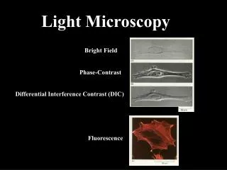
Today’s topics • Resolution • vertical and horizontal resolution limits • Different types of tip • Tip-related artefacts • Other artefacts • Streaks • Scanner bow • Tip changes • Periodic noise • Image processing and removal of artefacts • Available image processing software • Reducing and removing common artefacts • Image display and image enhancement Atomic force microscopy
Vertical resolution • The absolute limit of the vertical resolution is determined by the resolution of the vertical scanner movement which is <1Å. • Pixellation: • AFM data is recorded digitally. • The total number of available data points in the vertical direction can thus limit the vertical resolution. • The number of data points in the vertical dimension is determined by the conversion of a fixed number of bits over the full vertical range of the scanner. • Reducing the vertical range of the scanner can increase the sampling resolution in the vertical dimension to image sub-angstrom height changes. • In practice, the overall system noise is usually the primary factor limiting the vertical resolution: • Combined effects from electrical, mechanical, and acoustic sources. • Best achievable vertical resolution on departmental AFM: ca. 0.3 Å Atomic force microscopy
Pixellation Large vertical range Low sampling density in z Small vertical range High sampling density in z Atomic force microscopy
5 – 15 nm Lateral resolution • Lateral resolution is primarily determined by the radius of curvature of the end of the tip. • The sidewall angles of the tip will also determine its ability to probe high aspect ratio features. • Typical tip radii quoted by the manufacturers for tapping mode tips are around 5 – 15 nm, but these may increase quickly with tip wear. Atomic force microscopy
Lateral resolution and pixellation • The number of lines in an AFM scan, and the number of samples per line may be set in most AFM software. • Regardless of the pixel size, the feedback loop is sampling the topography many times at each pixel. • The data displayed at each pixel is the average of the sampling iterations by the feedback loop over the pixel area. • Obviously, features smaller than the pixel size will not be properly resolved. • Increasing the sampling density gives improved resolution but results in slower scanning. Atomic force microscopy
a w Dw 2 R w Dw 2 h h Errors in width measurements Assuming a hemispherical tip: h > R(1-cosα) R h < R(1-cosα) Not only the tip radius but the feature height and its likely shape must be known to accurately determine the true width. Atomic force microscopy
Blunt tip Sharp tip 200 nm 200 nm Dense nanostructure arrays For densely packed features the tip size can also cause errors in determining the height of the islands, or the overall appearance of the surface See animated illustration at: http://www.doitpoms.ac.uk/tlplib/afm/tip_related.php Atomic force microscopy
hemispherical tip R w h Terraced surfaces • Single atom-high steps may be routinely imaged by AFM. • However, as step-edges become closer together (e.g. on vicinal substrates) it may become impossible to see separate terraces. Atomic force microscopy
R dmeasured dpit a w Small pits • When imaging small pits, such as pits associated with threading dislocation terminations, one is unlikely to see the true pit depth. • The measured width will actually by more accurate than the measured depth, which will depend on the angle α, and the tip radius. • The pit will not be visible if the measured depth is less than the noise level. Atomic force microscopy
Images with very sharp “features” Given a realistic tip size these very sharp features cannot be real! Atomic force microscopy
Tip sidewall angles: Silicon nitride probes Atomic force microscopy
Tip sidewall angles: Etched silicon probes Atomic force microscopy
10° 90° 55° 73° 73° Tip sidewall angles and trench measurements Atomic force microscopy
Specialist tips: trench measurment Various high aspect ratio tip designs have been realised (usually using FIB) to try and overcome the trench measurement problem. Atomic force microscopy
z x “Critical dimension AFM” for trench measurement • “Critical dimension AFM” (CD-AFM) is a tool for the semiconductor fabrication industry for accurate trench measurement including assessment of sidewall roughness, overhangs etc. • In addition to the specialist tips shown, it requires additional instrumentation – providing tip oscillation and feedback in the x direction as well as the z direction, in order to image trench sidewall features. “Flared” or “boot-shaped” tips Atomic force microscopy
Specialist tips: improved nanostructure resolution (1) “Supersharpsilicon” probes employ nano-machining of standard tips to achieve a tip radius of less than 5 nm. They are very expensive and not at all robust Hi’Res probes are produced by the growth of diamond-like carbon whiskers on the ends of silicon tips. Sometimes several whiskers grow on one tip, leading to highly distorted images. Atomic force microscopy
Specialist tips: improved nanostructure resolution (2) • Tips with carbon nanotubes at the apex may be made by… • bringing tip close to a bundle of nanotubes, until one (or more) tubes sticks. • using nano-manipulators in a FEG-SEM. • Direct growth using an ethylene precursor and an iron catalyst. C.L. Cheung et al. PNAS, 97, 3809 (2000) Atomic force microscopy
Atomic resolution in AFM • Atomic resolution AFM not achievable in air due to effective broadening of tip by condensed water on surface. • Operating in fluid or under ultra high vacuum (UHV) solves this problem. • van der Waals forces operate over a much longer range than the tunnelling current in STM, so that for UHV operation, additional instrumentation aspects also had to be addressed before true atomic resolution could be achieved. From: Erlandsson R, Phys. Rev. B, 54, R8309 (1996) STM image of Si(111) Non-contact AFM image of Si(111) Atomic force microscopy
200 nm 200 nm Tip shape artefacts: damaged tip Normal tip Worn or damaged tip: several features have the same shape. In fact the small nanostructures are effectively imaging the tip shape, rather than vice versa. Atomic force microscopy
Damaged tip: more severe examples Atomic force microscopy
Tip shape artefacts: double tip Double or multiple tip images are formed when the tip has two or more end points which contact the sample while imaging. Individual features may appear sharp and small but may appear in pairs or larger groups. Atomic force microscopy
Hi’Res tips and multiple tip images Image of same sample taken using Hi’Res probe: this particular probe appears to have multiple whiskers involved in imaging. Image taken using normal Si probe Atomic force microscopy
Tip damage – scale effects Tips which give good images on flat surfaces may still show artefacts when much rougher features are imaged. Atomic force microscopy
Tip artefacts on large islands: example New tip Damaged tip Atomic force microscopy
Data from (a mostly excellent paper) So, what’s up with this image? A schematic, from the paper, based on this data: Atomic force microscopy
Checking for tip artefacts • For features with a clear orientation • Rotate the sample by 90°: if the features still have the same orientation as in the previous image, it’s a tip artefact. • Don’t be tempted to rotate the scan direction instead. This is NOT equivalent in this case. • Use a different tip. If the features now have a different shape, then the last tip was probably damaged. • If in doubt, try a third tip and go with the majority verdict! See animated illustration at: http://www.doitpoms.ac.uk/tlplib/afm/tip_related.php Atomic force microscopy
Sources of tip-shape change • Tip shapes may change during scanning due to: • Tip wear as the tip is rubbed over the surface • Particularly in contact mode if the material imaged is of similar hardness to the tip - e.g. GaN, diamond. • Damage if the tip hits a large surface feature without the feedback circuit having time to respond • This may occur when the scan rate is too fast over a rough surface, or when the feedback parameters have not been carefully set. • Picking up loose debris from the surface • Particularly if samples have been left around in air to gather dust or have been cleaved. Avoid this by dusting your samples with clean N2 before use. Atomic force microscopy
Scanner-related artefacts: Scanner bow • Because scanners are attached at one end and move the sample or tip on the other, the free end does not move in a level plane. • The mechanical properties of the piezo, as well as the kinematics of motion, may result in 2nd order or 3rd order deviations from an ideal plane. • This artefact is known as “bow”, and increases with scan size. Atomic force microscopy
Scanner-related artefacts: Scanner creep (1) • Creep is the drift of the piezo displacement after a DC offset voltage is applied to the piezo. • When a large voltage is applied, the scanner does not move the full distance required all at once. • It initially moves the majority of the distance quickly, and then slowly moves over the remainder. • If normal scanning resumes before the scanner has moved through the full distance then the image will be distorted by this residual movement. Atomic force microscopy
Scanner-related artefacts: Scanner creep (2) • Creep is likely to occur when… • …large changes in the X & Y offsets have been applied • …using the frame up and frame down commands, when the piezo travels over most of the scan area to restart the scan • …the scan size is changed abruptly by a large amount. X offset of 10 µm applied at this point Y X More examples at: http://www.doitpoms.ac.uk/tlplib/afm/scanner_related.php Atomic force microscopy
Resulting image (for retrace – i.e. right-to-left scan lines) First feature only impinges on tip for part of a scan line Tip misses second feature entirely Minimising the effects of creep and drift Atomic force microscopy
Resulting image (for retrace – i.e. right-to-left scan lines) Minimising the effects of creep and drift Although features are clearly distorted, both are visible, and the spacing between them is preserved. Always scan perpendicular to step edges and other long thin features where possible to minimise the impact of creep and drift on your image. This also makes image processing easier. Atomic force microscopy
Closed-loop scanners • To correct for scanner creep or drift and to minimise the impact of scanner non-linearities closed-loop scanners have been developed. • As well as using calibrated changes in voltage to adjust the scanner position, closed-loop scanners have an additional sensor (either optical or capacitive) which monitors the scanner’s position. • An additional feedback loop then adjusts the voltage applied to the scanner so that it reaches the desired position. • Closed-loop monitoring often increases the noise-level of the system, and hence is not advisable (or indeed necessary) for small scan sizes. • It can be very helpful, however, at large scan sizes. Atomic force microscopy
Tip change Tip change Tip changes Atomic force microscopy
Flattening to remove scanner bow and tip changes Free AFM image processing software available from: http://www.nanotec.es/products/wsxm/download.php Atomic force microscopy
What does the “flatten” function do? • For each line of the scan (in the fast scan direction), a least-squares-fit polynomial is calculated. • This polynomial fit is subtracted from the original line. • After flattening the average Z value of each scan line is equal to 0V. • The result is that height information in the slow-scan direction is removed. • The order of polynomial used my be chosen: • 0th order = horizontal line • 1st order = straight line • 2nd order = parabola • 3rd order = cubic Atomic force microscopy
Raw data + 200 nm 0th order fit Subtract fitted line Rescale - 200 nm Flatten function: Illustration 100 nm Surface profile to be measured: + 2.5 µm Scanner z-range - 2.5 µm • Most microscopes use 0th or 1st order flattening in real time to make it easier to understand the data whilst it is being recorded. • This is sometimes called “real-time plane-fitting”, although it actually uses a line-fitting function. • Even if data is displayed in a flattened format, it is usually recorded in a raw format, since flattening can result in loss of real information. Atomic force microscopy
Loss of information in the slow scan direction (1) Fast scan direction 0th order flatten Fast scan direction 0th order flatten Atomic force microscopy
0th order flatten Loss of information in the slow scan direction (2) Atomic force microscopy
Flattening non-uniform surfaces (1) 0th order flatten Atomic force microscopy
Flattening non-uniform surfaces (2) Simple flatten Flatten discarding regions Atomic force microscopy
Only flatten when necessary! As-recorded Simple flatten Flattening only adds false streaks to this image! Atomic force microscopy
Artefacts due to loose matter on surface Loose debris on the sample surface can cause loss of image resolution and can produce streaking in the image. Debris falls off tip, and resolution is recovered Scan direction Piece of debris attaches to tip – feature size increases Loss of resolution Skips and streaking Atomic force microscopy
Removing minor streaking (1) Atomic force microscopy
Removing minor streaking (2) Atomic force microscopy
Artefacts due to optical interference • Sometimes seen on highly reflective samples • Interference between light reflected from the tip and from the sample surface produces periodic oscillations. • This can usually be avoided by re-aligning the microscope so that the laser is more accurately centred on the cantilever. Atomic force microscopy
Periodic noise • Periodic noise is usually due to electrical or mechanical sources. • If you change the scan size, but maintain the same scan rate, periodic noise will maintain the same period. Atomic force microscopy
Fourier transform – for the removal of periodic noise (1) FFT Atomic force microscopy
Pass just these frequencies: Fourier transform – for the removal of periodic noise (2) Atomic force microscopy