Download
1 / 18
180 likes | 507 Vues
The Optic Pathway and the Optic Tract. RHB 340. The Path that the Image Takes is Divided into Two Parts. The Optic Path. The Optic Tract. The Optic Pathway. Begins at the optic nerve disk. The Optic Nerve. There are two optic nerves. One for the right eye and one for the left.

E N D
The Optic Pathway and the Optic Tract • RHB 340
The Path that the Image Takes is Divided into Two Parts The Optic Path The Optic Tract
The Optic Pathway • Begins at the optic nerve disk
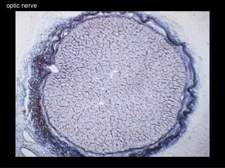
The Optic Nerve • There are two optic nerves. One for the right eye and one for the left. • The optic nerve attaches to the eye at the optic disc and runs back into the deeper parts of the brain. • Remember that it carries visual information encoded in electro-chemical signals.
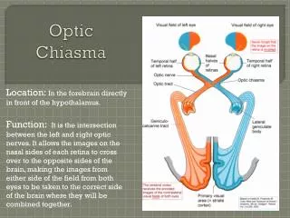
The Optic Chiasm • Impulses cross travel down the optic nerve and cross at the optic chiasm. • Some of the impulses, however, do not cross over to the opposite side of the brain. They continue down the same side.
The Optic Tract • After the chiasm, it becomes the optic tract. • The lateral geniculate bodies (sensory way stations) are the next big encounter for the visual image. • Here, the image begins to be integrated with other senses.
In the Optic Tract . . . • The fibers fan out into the visual cortex which is located at the top and back of the brain.
The Brain and Vision • Temporal Lobes • center for visual learning • recognition by sight • Midbrain -- Limbic sector • emotional responses to visual stimuli • Midbrain -- Superior Colliculus -- • guides visual attention
Some of the Visual Impulses are Processed in the Colliculus • These impulses are translated into what some researchers have termed “collicular vision.” • Collicular vision is sometimes called “blind sight.”
The person functions as a totally blind person unless they are moving through space. • Some people call collicular vision “travel vision.” • James E. Jan, MD, Peter K.H. Wong, MD, Maryke Groenveld, PhD, Olof Flodmark, MD, PhD, Creig S. Hoyt, MD