Download
1 / 9
120 likes | 379 Vues
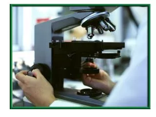
The use of microscope. Lab 3. Dissecting microscope. Low-power dissecting microscopes are better than hand lenses for examining colony morphology. Light Microscope.

E N D
The use of microscope Lab 3
Dissecting microscope • Low-power dissecting microscopes are better than hand lenses for examining colony morphology
Light Microscope • Some bacteria are so small that they are almost at the limit of resolution of the light microscope, while others are large enough to be readily visible. However, all are so small that they must be viewed with the oil immersion lens of the light microscope if their morphology is to be determined accurately.
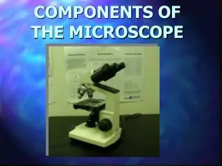
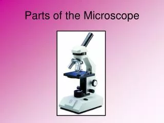
Light Microscope • 1. Ocular lens • 2. Objective turret • 3. Objective lens • 4. Coarse adjustment • 5. Fine adjustment • 6. Stage, to hold the sample • 7. Light source • 8. Condenser
Dark Ground Microscope • Organisms such as Leptospira are so thin that they are difficult to see by light microscopy even when stained. When direct light from below is blocked off in the dark ground condenser and directed at an angle through a wet preparation, particles of bacteria in its path are brightly illuminated against a black background
Phase Contrast Microscope • This system is used to observe features of unstained living cells or bacteria and involves condensers and lenses which cause the phase change in light passing through a transparent object to be translated into an image detectable by the eye as a lighter or darker region. Bacteria which interrupt the incident light in wet preparations appear as black objects against a light ground. Definition is better with this system than with dark ground microscopy as light scattering does not occur and a range of contrast is obtained.
Fluorescence Microscopy • In this system, ultraviolet light is directed through the specimen. Some bacteria possess autofluorescence and may be distinguished clearly, but more commonly specific bacteria are demonstrated by labelling with antibody attached to a fluorescent dye i.e. immunofluorescence. Recently antibodies have been linked to peroxidase and these can be used in ordinary light.
Electron Microscope • Standard techniques of electron microscopy are widely used in the study of ultrastructural features of bacteria. External features such as flagellae, fimbriae and cell wall surfaces are most frequently examined using negative staining techniques