Download
1 / 30
310 likes | 349 Vues
Restriction Enzyme Digestion of DNA. Experiment Goals. Digestion of DNA by restriction enzyme Analyze digested DNA by electrophoresis. Restriction Enzymes.

E N D
Experiment Goals • Digestion of DNA by restriction enzyme • Analyze digested DNA by electrophoresis
Restriction Enzymes • Restriction enzymes are endonucleases (Endo (inside), nuclease(cuts nucleic acid), which catalyze the cleavage of the phosphodiester bonds within both strands of DNA. • They require Mg+2 for activity and generate a 5 prime (5') phosphate and a 3 prime (3') hydroxyl group at the point of cleavage.
The distinguishing feature of restriction enzymes is that they only cut at very specific sequences of bases. This specific DNA sequence is called recognition sequence. Restriction enzymes are traditionally classified according to the subunit composition, cleavage position, sequence-specificity and cofactor requirements.
A restriction enzyme requires a specific double stranded recognition sequence of nucleotides to cut DNA. • Recognition sites are usually 4 to 8 base pairs in length. • Cleavage occurs within or near the site.
Naming of Restriction enzymes • Restriction enzymes are named according to the organism from which they are isolated. • This is done by using the first letter of the genus followed by the first two letters of the species and additional letter or number represent the strain or serotypes. • Only certain strains or sub-strains of a particular species may produce restriction enzymes.
Example of RE naming: HindII: Restriction enzyme was isolated from Haemophilus influenzae serotype d • There are hundreds of different REs from different microorganisms. • Different restriction enzymes have different recognition sequences. • This makes it possible to create a wide variety of different gene fragments.
Many recognition sequences are palindromic. For example, • 5’ GAATTC 3’ • 3’ CTTAAG 5’ palindromic: read the same in the opposite direction
Sticky and Blunt end cutters Not all restriction endonucleases cut symmetrically and leave blunt ends. Many endonucleases cleave the DNA backbones in positions that are not directly opposite each other or can make staggered cuts, which produce single stranded “sticky-ends” DNA from different sources can be spliced easily because of these sticky-end overhangs.
HaeIII Some restriction enzymes cut DNA at opposite base. They leave blunt ended DNA fragments These are called blunt end cutters
Restriction Enzyme Uses 1. Recombinant DNA technology *Discovery of enzymes that cut and paste DNA make genetic engineering possible. *Restriction enzyme cuts DNA and generates fragments *Ligase joins different DNA fragments *DNA fragments from different species can be ligated (joined) to create Recombinant DNA
2. Cloning • Replicates a sequence inserted into a host cell • 3. DNA restriction mapping The location of the restriction enzyme cleavage sites on the DNA molecule.
Biological Function • Restriction enzyme is part of the cell’s restriction-modification system in bacteria. • The phenomenon of restriction modification in bacteria is a small scale immune system for protection from infection by foreign DNA. • Bacteria can protect themselves only after foreign DNA has entered their cytoplasm.
Unit Determination Assay • One unit of restriction endonuclease is defined as the amount of enzyme required to digest one microgram of the appropriate substrate DNA completely in 60 minutes under the conditions specified for that enzyme.
Set up of a restriction enzyme reaction • A RE reaction contains the DNA to be analyzed, • A restriction enzyme, • A restriction enzyme buffer mix. • contains a buffering agent to maintain constant pH, • and Mg++ (from MgCl2) as a necessary cofactor for enzyme activity.
Digestion by Restriction Enzyme • Measure the DNA concentration • Use the Nano-drop spectrophotometer to measure the concentration of DNA, this is used to determine the amount of HinfI restriction enzyme to be used. Digestion of DNA • Mix the following components in a clean microtube. • Mix gently and spin down for a few seconds. • Incubate at 37°C for 16 hours.
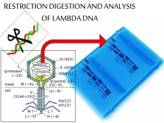
Analysis of DNA digestion • Analyze products on 2% agarose gel containing ethidium bromide. • Samples are prepared with loading dye and then loaded on the gel. • Visualize the PCR product on UV transilluminator.
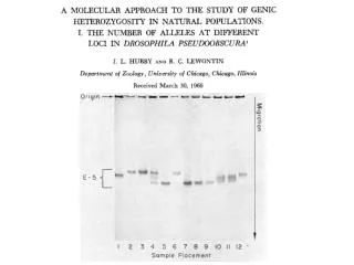
Electrophoresis of Genomic DNA Odd numbered lanes contain undigested genomic DNA Even numbered lanes contain digested genomic DNA Undigested DNA is represented by a sharp band near the wells of the gel, while smearing indicates digested DNA sample.
Q1Restriction enzyme AluI which recognize sequence5’ AGCT 3'3' TCGA 5‘ What are the double strand cut in DNA ( begin at G from 5’)
Q2: PstI RE recognize 5’ CTGCA*G 3' 3' G*ACGTC 5’ What are the digestion products?