Download
1 / 35
380 likes | 633 Vues
Restriction enzyme mapping. MISS: ISREAA F.HASSONA. Experiment Goals. Analysis of undigested plasmid on agarose gel electrophoresis for assessing plasmid preparation To perform restriction digestion of plasmid DNA Draw a plasmid map from gel electrophoresis data of the digested plasmid.

E N D
Restriction enzyme mapping MISS: ISREAA F.HASSONA
Experiment Goals Analysis of undigested plasmid on agarose gel electrophoresis for assessing plasmid preparation To perform restriction digestion of plasmid DNA Draw a plasmid map from gel electrophoresis data of the digested plasmid.
principle Agarose gel electrophoresis is a powerful separation method frequently used to analyze plasmid DNA. The microscopic pores present in agarose gels act as a molecular sieve. When an electric field is applied, the DNA molecules are separated by the pores in the gel according to their size and shape. The rate at which a given DNA molecule migrates through the gel depends not only on its size and shape, but also on the type of electrophoresis buffer, the gel concentration and the applied voltage.
Factors Affecting the Movement of DNA: Agarose Concentration: Higher concentrations of agarose facilite separation of small DNAs, while low agarose concentrations allow resolution of larger DNAs. The image shows migration of a set of DNA fragments in three concentrations of agarose, all of which electrophoresed at the same voltage and for identical times. Notice how the larger fragments are much better resolved in the 0.7% gel, while the small fragments separated best in 1.5% agarose
Voltage Applied As voltage increases, the speed of DNA also increases. But voltage should be limited because it heats and finally causes the gel to melt. Ethidium Bromide(EtBr) It is an intercalating agent which intercalates between nucleic acid bases and allows the convenient detection of DNA fragments in gel. When exposed to UV light, it will fluoresce with an orange colour. After the running of DNA through an EtBr-treated gel, any band containing more than ~20 ng DNA becomes distinctly visible under UV light.
Electrophoresis Buffer: Several different buffers have been recommended for electrophoresis of DNA. The most commonly used for duplex DNA are TAE (Tris-acetate-EDTA) and TBE (Tris-borate-EDTA). DNA fragments will migrate at somewhat different rates in these two buffers due to differences in ionic strength. Buffers not only establish a pH, but provide ions to support conductivity. Conversely, if you use concentrated buffer (e.g. a 10X stock solution), enough heat may be generated in the gel to melt it.
Conformation of DNA Plasmid DNA may appear in one of five conformations, which run at different speeds in a gel electrophoresis . Nicked open-circularDNA has one strand cut. Relaxed circularDNA is fully intact with both strands uncut, but has been enzymaticallyrelaxed(supercoils removed). LinearDNA has free ends because both strands have been cut . Supercoild orcovalently closed-circular) DNA is fully intact with both strands uncut, and with an integral twist, resulting in a compact form. SupercoileddenaturedDNA is likesupercoiled DNA, but has untwisted regions that make it slightly less compact; this can result from excessive alkalinity during plasmid preparation.
Conformation of Plasmid DNAs The relative electrophoretic mobility (speed) of these DNA conformations in a gel is as follows: Nicked Open Circular (slowest) Linear Relaxed Circular Supercoiled Denatured Supercoiled (fastest)
Supercoiled plasmid Supercoiling in the cell is caused by the action of enzymes called DNA gyrases. These enzymes use the chemical energy in ATP to introduce supercoiling into a relaxed molecule. In addition, there are enzymes that relax supercoiled DNA and are called unwinding or relaxing enzymes Topoisomerase. Supercoiling has important biological consequences. Very large DNA molecules would simply not fit in the cell if they were not supercoiled.
Supercoiled DNA has the fastest migration rate of the different forms of plasmid. • A small, compact supercoiled ccc-DNA sustains less friction against the agarose matrix than does other non-supercoild form.
Nicked open-circular Inthe laboratory, following a careful plasmid prep, most of the DNA will remain supercoiled, but a certain amount will sustain single-strand nicks. Given the presence of a break in only one of the strands, the DNA will remain circular, but the break will permit rotation around the phosphodiester backbone and the supercoils will be released.
This is because: Nicking can also be introduced by mechanical manipulations during plasmid purification.(or mechanical stress ). Overincubation during the alkaline lysis step. Total incubation of cell suspension with Cell Lysis Solution should not exceed 5 minutes. plasmids preps that have been thawed and refrozen many times, show more oc DNA than fresh preps. Endonucleases, such as DNAse I, will randomly nick supercoiled DNA .(enzymatic activity )
Linear plasmid Linearized DNA occurs when the DNA helix is cut in both strands at the same place. If you get linear DNA when you are hoping for supercoiled (e.g. after a plasmid prep) it is due to nuclease contamination or harsh treatment during purification . The linear DNA form is easily identified on an agarose gel by comparing uncut plasmid DNA with a sample of the plasmid that has been linearized using a restriction enzyme.
Linear DNA runs through a gel sustains less friction than open-circular DNA, but more than supercoiled. (faster than nicked).
Catenanes: During replication, several plasmid molecules can form interlocking rings with themselves. These forms are called catenanes. Catenanes can contain 2 plasmid molecules (dimer) Three molecules (trimer), etc. (Figure 7).
Catenanes migrate more slowly than single circular DNA that are nicked during electrophoresis. Dimers migrate faster than trimers, which migrate faster than tetramers, etc. Catenanes give rise to the same final restriction enzyme cleavage patterns as their uncatenated (single circular) forms.
Note: Circular, single stranded plasmid During alkaline lysis, plasmids are denatured because the hydrogen bonds are disrupted by the alkaline conditions. But the covalently-closed circular strands remain intact and topologically constrained and when the pH is returned to neutral the hydrogen bonds reform and the supercoiled DNA is re-formed. However, if the alkaline lysis step is overly harsh (e.g. it is incubated for too long) the DNA can become permanently denatured and give you useless single stranded closed circles that migrate ahead of all of the other forms of the plasmid in a gel.
NOTE If RNA contamination is present, one would see a faint and smeary RNA band below the supercoild as RNA contamination is observed when the RNase treatment has not been carried out properly.
Uncut plasmids Uncut plasmids will be run as a control – no restriction enzyme added. Running these controls allows us to know which bands in gel are due to uncut plasmids.
Uncut Plasmid Forms Uncut plasmid are most likely to exist in one of 3 forms: Supercoiled Nicked Multimer
Uncut Plasmids on a Gel When you run the uncut plasmids on a gel, you will get a pattern of bands (at least 3-4) Which band of DNA is the supercoiled plasmids? Nicked circles? Multimers? - Multimer supercoiled Nicked circle +
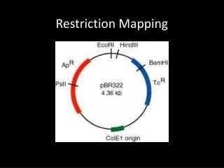
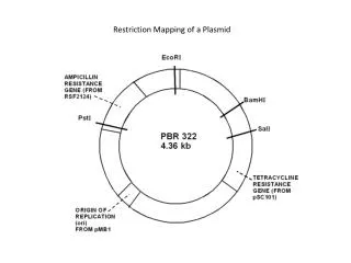
BACKGROUND • Mapping of DNA restriction sites is an important part of working in a molecular biotechnology. • TO find out : • Which restriction enzymes will cut a particular plasmid • How many places a restriction enzyme will cut a particular plasmid • How far apart restriction sites are.
Restriction enzyme mapping determines the relative positions of cleavage sites to one another in a DNA molecule. This is done by determining sizes of DNA fragments generated by different combinations of restriction enzyme digests.
As an example, suppose that you are working with a 4,000 bp (4 kb) plasmid (a small circular piece of DNA) that has restriction sites for EcoR I, BamH I, and Hind III. When you cut the plasmid with any one of the enzymes, and run each digestion on an agarose gel, you see that one band of DNA is present and runs the same distance as the 4 kb fragment in the standard marker DNA.
These data show that each enzyme has only one restriction site within the plasmid. Without further experimentation, there is no way to know where each of the three sites is located with respect to the other two. Therefore, double and even triple digestions are performed, run on a gel, and analyzed.
Example 2, consider a 5000 base pair, circular plasmid DNA containing single recognition sites for enzymes A, B, and C. Any one of these enzymes will cleave the DNA once to produce a linear molecule of 5000 base pairs. Differently paired combinations of enzymes in the same reaction mixture (double-digests) will produce the following DNA fragments (sizes in base pairs):
The triple digest, A + B + C is a confirmatory test
Running Plasmid DNA on an agarose gel • 1- Prepare Samples: Prepare 1ug of plasmid DNA in 1X load dye in a 1.5mL microcentrifuge tube. For example: DNA yield is 100ng/ul = 10ul plasmid DNA 10X load dye = 1 ul of 10X load dye Alternatively, prepare 20ul of the eluted DNA in 1X final concentration of load dye. • 2-Prepare molecular weight Marker: Prepare 1ul of 1Kb DNA Step Ladder molecular weight marker in 1x load dye in a 1.5mL microcentrifuge tube as follows: 1ul 1Kb DNA Step Ladder 8ul dH2O 1ul 10X load dye BIOL1414 Lab Manual Fall 2011 138
. Save the remaining plasmid DNA in a labeled microcentrifuge tube for the next lab exercise. Plasmid DNA should be stored at 4oC. • 4. Load sample in load dye into one well and DNA ladder in load dye into another well. • 5. Run at 100 volts for approximately 30 minutes (until the dye is approximately 1cm from the bottom of the gel). Record loading position in the gel electrophoresis documentation form. • 6. Visualize DNA bands by placing gels on a UV transilluminator and capture an image using the gel electrophoresis documentation system. Affix a well-labeled copy of your gel picture to the gel electrophoresis documentation form and turn this in with your lab