Download
1 / 1
10 likes | 177 Vues
TIMC Laboratory Université Joseph Fourier GRENOBLE FRANCE . Laboratoire de biomécanique TOULOUSE, FRANCE. 3D FINITE ELEMENT MESHING OF ENTIRE FEMORA BY USING THE MESH MATCHING ALGORITHM V. Luboz, B. Couteau, and Y. Payan Contact: Yohan.Payan@imag.fr. Abstract.

E N D
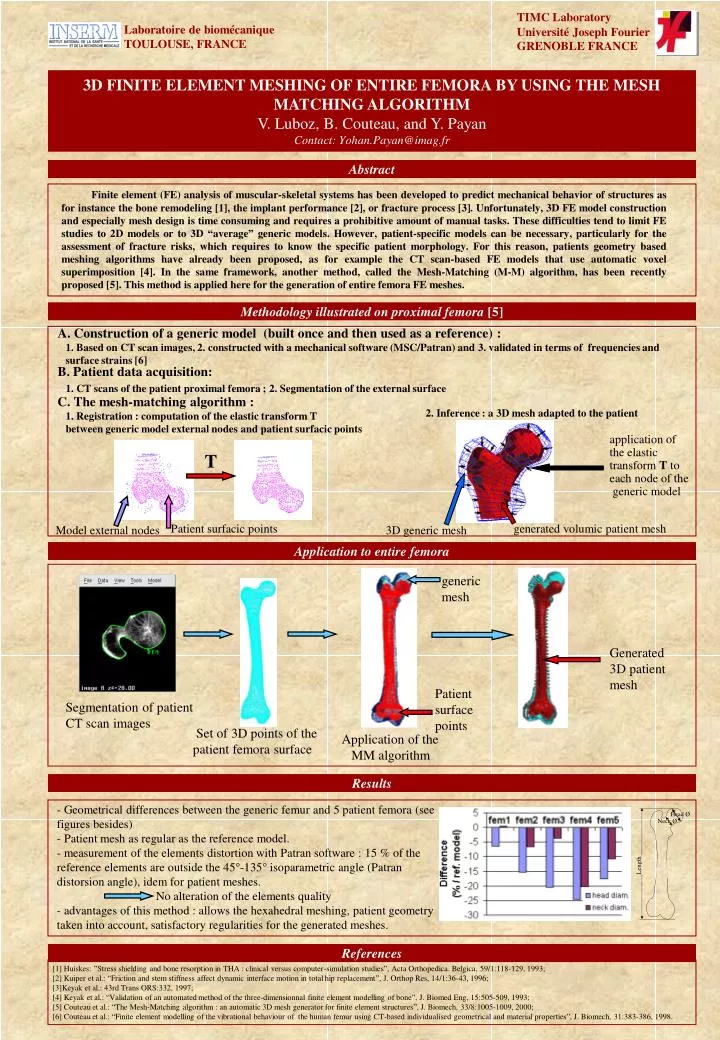
TIMC Laboratory Université Joseph Fourier GRENOBLE FRANCE Laboratoire de biomécanique TOULOUSE, FRANCE 3D FINITE ELEMENT MESHING OF ENTIRE FEMORA BY USING THE MESH MATCHING ALGORITHM V. Luboz, B. Couteau, and Y. Payan Contact: Yohan.Payan@imag.fr Abstract Finite element (FE) analysis of muscular-skeletal systems has been developed to predict mechanical behavior of structures as for instance the bone remodeling [1], the implant performance [2], or fracture process [3]. Unfortunately, 3D FE model construction and especially mesh design is time consuming and requires a prohibitive amount of manual tasks. These difficulties tend to limit FE studies to 2D models or to 3D “average” generic models. However, patient-specific models can be necessary, particularly for the assessment of fracture risks, which requires to know the specific patient morphology. For this reason, patients geometry based meshing algorithms have already been proposed, as for example the CT scan-based FE models that use automatic voxel superimposition [4]. In the same framework, another method, called the Mesh-Matching (M-M) algorithm, has been recently proposed [5]. This method is applied here for the generation of entire femora FE meshes. Methodology illustrated on proximal femora [5] A. Construction of a generic model (built once and then used as a reference) : 1. Based on CT scan images, 2. constructed with a mechanical software (MSC/Patran) and 3. validated in terms of frequencies and surface strains [6] B. Patient data acquisition: 1. CT scans of the patient proximal femora ; 2. Segmentation of the external surface C. The mesh-matching algorithm : 2. Inference : a 3D mesh adapted to the patient 1. Registration : computation of the elastic transform T between generic model external nodes and patient surfacic points application of the elastic transform T to each node of the generic model T Patient surfacic points generated volumic patient mesh Model external nodes 3D generic mesh Application to entire femora generic mesh Generated 3D patient mesh Patient surface points Segmentation of patient CT scan images Set of 3D points of the patient femora surface Application of the MM algorithm Results - Geometrical differences between the generic femur and 5 patient femora (see figures besides) - Patient mesh as regular as the reference model. - measurement of the elements distortion with Patran software : 15 % of the reference elements are outside the 45°-135° isoparametric angle (Patran distorsion angle), idem for patient meshes. No alteration of the elements quality - advantages of this method : allows the hexahedral meshing, patient geometry taken into account, satisfactory regularities for the generated meshes. References [1] Huiskes: ”Stress shielding and bone resorption in THA : clinical versus computer-simulation studies”, Acta Orthopedica. Belgica, 59/1:118-129, 1993; [2] Kuiper et al.: “Friction and stem stiffness affect dynamic interface motion in total hip replacement”, J. Orthop Res, 14/1:36-43, 1996; [3]Keyak et al.: 43rd Trans ORS:332, 1997; [4] Keyak et al.: “Validation of an automated method of the three-dimensionnal finite element modelling of bone”, J. Biomed Eng, 15:505-509, 1993; [5] Couteau et al.: “The Mesh-Matching algorithm : an automatic 3D mesh generator for finite element structures”, J. Biomech, 33/8:1005-1009, 2000; [6] Couteau et al.: “Finite element modelling of the vibrational behaviour of the human femur using CT-based individualised geometrical and material properties”, J. Biomech, 31:383-386, 1998.