Download
1 / 61
610 likes | 1.2k Vues
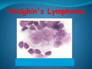
LYMPHOMA. Dr. Eyad F. Alsaeed Associate Professor Consultant Radiation Oncologist Department Of Medicine. Myeloid. Lymphoid. Histiocytic. Mast Cell. WHO Classification of Hematological Neoplasms. B cell neoplasms * T cell neoplasms Hodgkin’s lymphoma. * Includes plasma cell myeloma.

E N D
LYMPHOMA Dr. Eyad F. Alsaeed Associate Professor Consultant Radiation Oncologist Department Of Medicine
Myeloid Lymphoid Histiocytic Mast Cell WHO Classification of Hematological Neoplasms B cell neoplasms * T cell neoplasms Hodgkin’s lymphoma *Includes plasma cell myeloma
Proposed WHO Classification of. Lymphoid Neoplasms - 1 B-Cell neoplasms Precursor B-cell neoplasm Precursor B-lymphoblastic leukemia/Iymphoma (precursor B-cell acute lymphoblastic leukemia) Mature (peripheral) B-cell neoplasm* B-cell chronic lymphocytic leukemia/small lymphocytic lymphoma B-cell prolymphocytic leukemia Lymphoplasmacytic lymphoma Splenic marginal zone B-cell lymphoma (+/— villous lymphocytes) Hairy cell leukemia Plasma cell myeloma/plasmacytoma Extranodal marginal zone B-cell lymphama of MALT type Nodal marginal zone B-cell lymphoma (+1— monocytoid B cells) Follicular lymphoma Mantle-cell lymphoma Diffuse large B-cell lymphama Mediastinal large B-cell lymphoma Primary effusion lymphoma Burkitt’s lymphoma/Burlcitt cell leukemia
Proposed WHO Classification of. Lymphoid Neoplasms (cont’d) T-cell and NK-cell neoplasms Precursor T-cell neoplasm Precursor T-lymnphoblastic lymphoma/leukemia (precursor T-cell acute lymphoblastic leukemia) Mature (peripheral) T-cell neoplasms T-cell prolymphocytic leukemia T-cell granular lymphocytic leukemia Aggressive NK-cell leukemia Adult T-cell lymphoma/leukemia (HTLV1 +) Extranodal NK/T-cell lymphoma, nasal type Enteropathy-type T-cell lymphoma Hepotosplenic gamma-delta T-cell lymphoma Subcutaneous panniculitis-like T-cell lymphoma Mycosis fungoides/Sezary syndrome Anaplastic large-cell lymphoma, T/null cell, primary cutaneous type Peripheral T-cell lymphoma, not otherwise characterized Angioimmunoblastic T-celllymphoma Amaplastic large-cell lymphoma, T/null cell, primary systemic type Hodgkin’s lymphoma (Hodgkin’s disease) Nodular lymphocyte-predominant Hodgkin’s )ymphoma Classical Hodgkin’s lymphoma Nodular sclerosis Hodgkin’s Iymphoma (grades 1 and 2) Lymphocyte-rich classical Hodgkin’s lymphoma Mixed cellularity Hodgkin’s lymphoma Lymphocyte depletion Hodgkin’s lymphoma NOTE: Only major categories are included. Subtypes and variants will be discussed in the WHO book2 and are listed in Tables 7 through 16. Common entities are shown in boldface type. Abbreviations: HTLV1 +, human T-cell leukemia virus; MALT, mucosa-associated lymphoid tissue; NK, natural killer. *B-and T-/NK-cell neoplasms are grouped according to major clinical presentations (predominantly disseminated/leukemic, primary extranodal, predominantly nodal).
Clinical Grouping of LymphomasNHL • Indolent • Aggressive • Highly aggressive • Formerly • Low Grade • Intermediate Grade • High Grade
Clinical Grouping of Lymphomas Approximate International Incidence • Indolent( “low grade”) • Follicular lymphoma Grade 1,2 22% • Marginal zone lymphoma • Nodal 1% • Extranodal (MALT) 5% • Small lymphocytic lymphoma 6% • Lymphoplasmacytic* 1% *association with Waldenstrom’s macroglobulinemia
Clinical Grouping of Lymphomas Approximate International Incidence • Aggressive( “intermediate grade”) • Diffuse large B-cell lymphoma 21% • Primary mediastinal large B cell lymphoma 2% • Anaplastic large T / null cell lymphoma 2% • Peripheral T cell lymphoma 6% • Extranodal NK / T cell lymphoma, nasal type • Follicular lymphoma Gd 3 • Mantle cell lymphoma 6%
Clinical Grouping of Lymphomas • Highly Aggressive ( ”High grade”) • Lymphoblastic lymphoma 2% • Burkitts lymphoma 1% • Burkitt-like lymphoma 2% Approximate International Incidence
Clinical Grouping of Lymphomas(further simplified for radiation oncology exam purposes) • INDOLENT • Follicular lymphoma Gd 1, 2 • MALT (marginal zone lymphoma, extranodal (MALT type)) • AGGRESSIVE • Diffuse large cell
Lymphoma – Staging System(Cotswold’s Meeting modification of Ann Arbour Classification ISingle lymph node region (or lymphoid structure) * II2 or more lymph node regions III Lymph node regions on both sides of diaphragm IV Extensive extranodal disease (more extensive then “E”)
Lymphoma – Staging SystemSubscripts A Asymptomatic B Fever > 38o, recurrent Night sweats drenching, recurrent Weight loss > 10% body wt in 6 mos X Bulky disease > 10 cm or > 1/3 internal transverse diameter @ T5/6 on PA CXR ELimited extranodal extension from adjacent nodal site
Lymphoma – Essential Staging Investigations • Biopsy – pathology review • History – B symptoms, PS • Physical Exam – nodes, liver, spleen, oropharynx • CBC • creatinine, liver function tests, LDH, calcium • Bone marrow aspiration & biopsy • CT neck, thorax, abdomen, pelvis
for gastric lymphoma Additional Staging Investigations • PET or 67Ga scan • CT / MRI of head & neck • Cytology of effusions, ascites • Endoscopy • Endoscopic U/S • MRI - CNS, bone, head & neck presentation • HIV • CSF cytology - testis, paranasal sinus, peri-orbital, paravertebral, CNS, epidural, stage IV with bone marrow involvement
International Prognostic Index for NHL *Diffuse large cell lymphoma
Indolent Lymphomae.g. Follicular Gd 1/2, small lymphocytic, marginal zone Limited Disease ( Stage 1A, 2A if 3 or less adjacent node regions) • IFRT* 30-35 Gy • Expect ~ 40% long term FFR • Alternate: • CMT • Observation. Treat when symptomatic. *Involved Field Radiotherapy. Use 35 Gy for follicular. 30 Gy for SLL, marginal
Indolent Lymphomae.g. Follicular Gd 1/2, small lymphocytic, marginal zone Advanced Stage ( some Stage 2, Stage 3, 4 ) • Palliative RT* for localized symptomatic disease • Palliative chemotherapy** for disseminated symptomatic disease • Observation only if low bulk, asymptomatic • Treat when symptomatic * IFRT 15 – 20 Gy / 5 ** CVP, chlorambucil
Aggressive Lymphoma(e.g. Diffuse large B cell) Stage I, some Stage II • CHOP* x 3 + IFRT (35-45 Gy)** Expect ~ 75% long term FFR Stage III, IV, B symptoms, or bulky disease CHOP* x 6-8 IFRT (35-45 Gy) to - sites of initial bulk - residual disease (i.e. PR) *or CHOP-R (see next slide) ** higher radiation dose if residual disease
Aggressive Lymphoma(e.g. Diffuse large B cell) CHOP q 21 days • Cyclophosphamide • doxorubicin (formerly Hydroxydaunorubicin) • vincristine (“Oncovin”) • Prednisone (p.o. x 5 days) • CHOP-R x 8 ~40 % 3 yr EFS, OS (vs. CHOP x 8)
Extranodal Lymphoma • Same treatment as nodal lymphoma Notable Exceptions: • Gastric MALT • Testis • CNS • Skin
MALT = “mucosa associated lymphoid tissue” • MALT Lymphoma • Marginal zone B-cell lymphoma of extranodal (MALT) type • Stomach. assoc. with Helicobacter pylori infection* • Salivary Gland. assoc with Sjogren’s syndrome* • Thyroid. assoc with Hashimoto’s thyroiditis* • Orbital (lacrimal, conjunctiva) • Other: Waldeyer’s ring, breast, bladder, lung, skin * chronic antigen stimulation
Gastric MALT Lymphoma • Stage IE , H. pylori + • PPI, 2 antibiotics (e.g. clarithromycin, amoxicillin) F/U gastroscopy + Bx q6mo for 2 yrs, then q1yr • Stage IE, H. pylori - or antibiotic failure • IFRT 30 Gy (95% local control) • Stage 2 or higher • Treat as indolent lymphoma + H. pylori eradication
WHO Classification of Lymphoid Neoplasms Hodgkin’s Lymphoma ( Hodgkin’s disease) • Nodular lymphocyte-predominant HL* • Classical HL • Nodular sclerosis HL • Lymphocyte-rich classical HL* • Mixed cellularity HL • Lymphocyte depletion HL *formerly, both of these were classified as lymphocyte predominance Hodgkin’s Disease
Hodgkin’s Disease - Staging Investigations • Biopsy – pathology review • History – B symptoms, pruritis, alcohol pain, PS • Physical Exam – nodes, liver, spleen, oropharynx • CBC, ESR • creatinine, liver function tests, LDH, calcium, albumin • Bone marrow aspiration & biopsy • if abnormal CBC, Stage 2B or higher • CT thorax, abdomen, pelvis
Hodgkin’s Disease - Other Investigations • PET scan • 67Ga scan • Lymphangiogram – if expertise available, no PET • Pregnancy test • oophoropexy / semen cryopreservation • if chemotherapy or pelvic RT • Dental assessment – if oropharyngeal RT
Hodgkin’s Lymphoma Early Stage 1A, 2A Advanced III, IV Bulky Disease B Symptoms
Hodgkin’s Lymphoma Early Stage 1A, 2A Advanced III, IV Bulky Disease B Symptoms • UNFAVOURABLE* • > 3 sites • Age > 40 • ESR > 50 • Mixed cellularity • FAVOURABLE* • 1-3 sites • Age 40 • ESR < 50 • NS, LRCHL *NCIC HD6 Study Criteria reflecting prognosis when treated with radiation only
Early Stage Hodgkin’s LymphomaFavourable Prognosis • ABVD X 3 - 4 • IFRT 30 Gy / 20 • Fewer cycles ABVD may be adequate. GHSG HD10 study, in progress, compares ABVD x 2 vs. ABVD x 4 • Lower radiation dose may be adequate. GHSG HD10 study and EORTC H9 study, in progress, compare IFRT 20 Gy with 30 Gy (HD10) and 36 Gy (H9) • Caution:late toxicity data awaited
STNI ABVD x 2 + IFRT ABVD x 6 historical gold standard survival CMT use if CTx containdicated but: high risk late toxicity as per BCCA guidelines awaiting clinical trial results (GHSG HD10) awaiting NCIC HD.6 results Favourable Prognosis – Early Stage Hodgkin’s LymphomaSome Other Treatment Options Mantle + Para-aortic nodes,spleen 35 Gy/20
Early Stage Hodgkin’s LymphomaUnfavourable Prognosis • ABVD X 4 - 6 • IFRT 30 Gy / 20 • NB: Overlap with favourable prognosis ESHL
Advanced Stage Hodgkin’s LymphomaStage 3, 4, B symptoms, bulky disease • ABVD X 6 – 8* • IFRT • sites of bulky disease • sites of residual disease (35 Gy / 20) * ABVD until 2 cycles past maximum response
ABVD • doxorubicin (Adriamycin) • Bleomycin • Vinblastine • Dacarbazine IV Days 1, 15
Very Favourable Prognosis Hodgkin’s Lymphoma • Stage 1A NLPHL* • Stage 1A high neck NS, LRCHL IFRT 35 Gy / 20 • *Nodular Lymphocyte Predominant HL • usually localized, peripheral nodal sites • good prognosis, but some late relapses (>10yr)
Hodgkin’s LymphomaRough Approximation of Prognosis If RT only (STNI): Deaths from 2nd malignancy > deaths from Hodgkin’s disease by 15 – 20 yrs * Depending on Hasenclever Prognostic Index: based on Age>45, male, Stage 4, albumin < 4, Hb < 10.5, WBC<600 or >15000
Side Effects of Radiotherapy for Hodgkin’s Lymphoma • Depend on • Dose/fractionation • Site • Irradiated volume • Chemotherapy • - Acute - Subacute - Late
Toxicity of STNI for Hodgkin’s Lymphoma ACUTE • Skin erythema • Local alopecia • Xerostomia • Dysphagia • Fatigue • WBC, platelets • Para-aortic RT - nausea, vomiting - diarrhea
Toxicity of EFRT for Hodgkin’s Lymphoma SUBACUTE • Fatigue • Xerostomia • Pneumonitis < 5%, dependent on lung volume treated • Herpes Zoster • Lhermitte’s Syndrome
Toxicity of STNI for Hodgkin’s Lymphoma LATE • Hypothyroidism • Cardiac • (CAD, valvular disease, pericarditis) • 5% risk cardiac death in 20 yrs (2-3 x expected) • 2nd malignancy ( risk of most solid tumors) • esp. breast ca if < 25 yrs at time of RT • Lung ca in smokers • Solid tumour risk rises after 10 years from RT • Absolute Excess Risk ~1% per year
52 y.o. male with dysphagia Exam: posterior oropharyngeal mass involving L tonsil, L base of tongue, crossing over midline to involve R base of tongue. Biopsy: “large cell lymphoma of T-cell derivation with differential diagnosis between nasal type extranodal T-cell lymphoma, and peripheral T-cell lymphoma of unspecified type.”
General Principles of Answering Lymphoma Questions - 2 • “First of all, I would take a complete history and perform a full physical examination…” • “The pathology should be reviewed by an experienced lymphoma pathologist…” • “This patient’s management should be discussed in a multidisciplinary setting*…” *At least by haematologist / medical oncologist and radiation oncologist
Clinical Grouping of Lymphomas Approximate International Incidence • Aggressive( “intermediate grade”) • Diffuse large B-cell lymphoma 21% • Primary mediastinal large B cell lymphoma 2% • Anaplastic large T / null cell lymphoma 2% • Peripheral T cell lymphoma 6% • Extranodal NK / T cell lymphoma, nasal type • Follicular lymphoma Gd 3 • Mantle cell lymphoma 6%
“Aggressive” lymphoma • CT head, neck, thorax, abdo, pelvis • MRI head & neck • CBC, creatinine, LDH, liver enzymes • Bone marrow aspiration & biopsy • HIV testing • Dental consult
DM 005676 CT: “nodular defect arising from posterior aspect of pharynx extending into tonsillar region…3.5 x 1.5 cm…also a prominent nodular structure extending through base of tongue 3.5 x 2.5 cm…. Non-specific cervical lymph nodes, the largest 11 mm…” No evidence of disease at other sites, normal lab work.
DM 005676 CHOP x 3 Why not CHOP-R? Planning CT Supine, in immobilization shell GTV contoured
DM 005676 PTV: Waldeyer’s Ring. Lateral POP, 6 MV photons, compensators for dose homogeneity, 40 Gy / 20 / 4 wks
31 y.o. female with recent onset fatigue, night sweats, and mass in right neck Seen in ER: R supraclavicular node ~2 cm CXR: Huge ant mediastinal mass Biopsy: Nodular sclerosis type Hodgkin’s disease CT Chest: “Large, lobulated mass in anterior mediastinum extending from suprasternal notch to cardiophrenic angle…also an enlarged subcarinal node…”
Referral to Radiation Oncology • History & Physical • Pathology Review • Discuss with Haematologist / Medical Oncologist • CBC, ESR, creatinine, liver enzymes • CT abdo-pelvis • 67Ga scan • Bone marrow aspiration & biopsy
BW 037843 Hodgkin’s Lymphoma, Nodular Sclerosis type Stage IXB