Download
1 / 13
140 likes | 432 Vues
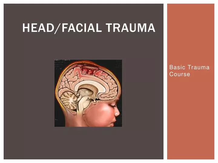
Head/Facial Trauma. Basic Trauma Course. Mechanism of Injury. Head injuries are most often caused by Motor Vehicle Crashes (MVC), especially in teens and young adults. Falls are the next most common cause, but falls tend to occur most in the extremes of age – the very young and the very old.

E N D
Head/Facial Trauma Basic Trauma Course
Mechanism of Injury • Head injuries are most often caused by Motor Vehicle Crashes (MVC), especially in teens and young adults. • Falls are the next most common cause, but falls tend to occur most in the extremes of age – the very young and the very old. • Alcohol and other drug use also play a significant factor in many head injuries.
Injury to the Brain • The “Primary Insult” refers to the initial traumatic damage to the brain. • “Secondary Insult” is any development that contributes to the neurologic injury and occurs subsequent to the initial injury. • Treatment is now focused on minimizing or avoiding secondary injury. • Some examples of secondary insults are hypotension, increased ICP, decreases in blood oxygen, increases in oxygen demand, increases in body temperature, increased blood glucose levels, and electrolyte abnormalities.
Airway Management • Airway obstruction caused by occlusion by the tongue, accumulation of secretions or blood, and/or facial edema is the main cause of death in brain and craniofacial trauma. • Hyperventilation????? • PaCO2 levels between 35-40 are ideal; PaO2 should be 90 or higher. PEEP has been found to affect ICP at levels over 10 cm H2O, but is not harmful at levels up to 10 of PEEP. • Early intubation is generally suggested in moderate to severely injured patients.
Fluid Status Management • We strive to achieve normovolemia.. • Fluid resuscitate a patient • Place them on low dose Dopamine to increase mean arterial pressure (MAP) • Administer Mannitol
ICP • Intracranial pressure is a reflection of three volumes: Brain, CSF, and blood within the nonexpansible cranial vault. • Normal ICP is 0-15. • ICP is often managed by the combined use of sedation (analgesics, benzodiazepines), CSF drainage via ventriculostomy, and Mannitol. • Mannitol is an osmotic diuretic used to dehydrate the brain, not the entire body. • Herniation occurs as a result of uncontrolled increases in ICP. Symptoms include: Unilateral or bilateral pupillary dilation, asymmetric pupillary reactivity, abnormal motor posturing, or other evidence of neurologic deterioration.
Controlling ICP • Measures to control or treat intracranial pressure: • Prevent herniation, if possible • Maintain patent airway • Normalize blood gases and electrolytes • Control seizures (may increase ICP and increase O2 / glucose demand) – phenytoin is preferable to benzodiazepines to minimize sedation and apnea • Maintain head of bed to 30 degrees, head position midline • Normalize temperature – cooling blankets may be necessary as Tylenol and Motrin are not effective if the temp is neurologic in origin • Maintain euvolemia with isotonic IV solutions (lactated ringers or normal saline) • Mannitol 0.25 – 0.50 gm/kg over 15-30 minutes may be used to temporarily decrease ICP. Monitoring serum osmolarity and serum sodium levels are crucial with Mannitol therapy.
Diffuse Brain Injury • Concussion- Transient, reversible alteration in brain function. • Classic presentation includes transient loss of consciousness, with return to GCS of 14-15, often with repetitive questions and amnesia of the crash /event. Brain CT is negative. • Diffuse Axonal Injury-Impaired function and gradual loss of axons. This is often diffuse and not easily seen on CT- MRI often confirms diagnoses. If enough axons are injured, the ability of nerve cells to communicate is lost, resulting in severe deficits. Can be caused by hypoxic insults to the brain by prolonged shock or apnea after initial trauma.
Skull Fractures • Fragments depressed more than the thickness of the skull require surgical elevation • Complications • Infections • Hematoma • CSF leaks • Loss of smell • Loss of hearing • Seizures • Pneumocephalus Monitor for seizures, Monitor for CSF leak • Avoid nasal intubation, nasal gastric tube, nose blowing, sneezing
Focal Brain Injuries • Cerebral Contusion-Bruising of the brain surface with varying degrees of edema, usually associated with a countra-coup injury. Due to an increase in the edema a follow up CT may be worse with Blossoming of the edema. • Epidural Hematoma- A collection of blood between the skull and the dura mater. Associated with fractures of the temporal or parietal skull that lacerate the middle meningeal artery. • Subdural Hematoma- Hematoma beneath the dura mater that is venous in origin, usually as a result of acceleration, deceleration or a combination of both forces.
Ongoing Assessment • A thorough neurological exam is essential to adequate management of the severely head injured. • Subtle changes in exam may indicate an increase in the size of a mass lesion, the expansion of a contusion, or impending herniation. • If a change in neuro exam occurs, it is up to the bedside nurse to notify the physician and take measures to optimize ICP and hemodynamics until the physician arrives or orders are received. • Early detection and intervention are key in optimizing neurologic outcome.
Facial Trauma • Airway • Occlusion or compromise of the airway often results from facial trauma. Edema, blood, vomitus, and teeth can occlude the airway. • Bleeding • Facial injuries bleed profusely because of their vascular nature. Usually venous unless in the temporal area then it can be arterial in nature. • Neuro • Injury to the cranial nerves are the risk with facial injuries. Arterial injury can cause sensory impairment due to cerebral ischemia.
Eye Injury • Eye Injury • Eye injury can occur with periorbital or midface fractures. Injuries such as orbital fractures are almost always due to a blunt blow from a relatively large object. Entrapment of cranial nerves and muscles may produce the signs associated with this injury.