Download
1 / 42
420 likes | 550 Vues
Chapter 11: Nervous Tissue. The Nervous System. The master controlling and communicating system of the body Functions: Sensory input – monitoring stimuli occurring inside and outside the body Integration – interpretation of sensory input

E N D
The Nervous System • The master controlling and communicating system of the body • Functions: • Sensory input – monitoring stimuli occurring inside and outside the body • Integration – interpretation of sensory input • Motor output – response to stimuli by activating effector organs
Organization of the Nervous System • Central nervous system (CNS) • Brain and spinal cord • Integration and command center • Peripheral nervous system (PNS) • Paired spinal and cranial nerves • Carries messages to and from the spinal cord and brain
Peripheral Nervous System: Two Functional Divisions • Sensory (afferent) division • Sensory afferent fibers – carry impulses from skin, skeletal muscles, and joints to the brain • Visceral afferent fibers – transmit impulses from visceral organs to the brain • Motor (efferent) division • Transmits impulses from the CNS to effector organs
Motor Division: Two Main Parts • Somatic nervous system • Conscious control of skeletal muscles • Autonomic nervous system (ANS) • Regulate smooth muscle, cardiac muscle, and glands • Divisions – sympathetic and parasympathetic
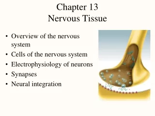
Histology of Nervous Tissue • The two principal cell types of the nervous system are: • Neurons – excitable cells that transmit electrical signals • Supporting cells – cells that surround and wrap neurons
Supporting Cells: Neuroglia • The supporting cells (neuroglia or glia): • Provide a supportive scaffolding for neurons • Segregate and insulate neurons • Guide young neurons to the proper connections • Promote health and growth
Astrocytes • Most abundant, versatile, and highly branched glial cells • They cling to neurons and cover capillaries • Functionally, they: • Support and brace neurons • Anchor neurons to their nutrient supplies • Guide migration of young neurons • Control the chemical environment
Microglia and Ependymal Cells • Microglia – small, ovoid cells with spiny processes • Phagocytes that monitor the health of neurons • Ependymal cells – squamous- to columnar-shaped cells • They line the central cavities of the brain and spinal column
Oligodendrocytes, Schwann Cells, and Satellite Cells • Oligodendrocytes – branched cells that wrap CNS nerve fibers • Schwann cells (neurolemmocytes) – surround fibers of the PNS • Satellite cells surround neuron cell bodies with ganglia
Neurons (Nerve Cells) • Structural units of the nervous system • Composed of a body, axon, and dendrites • Long-lived, amitotic, and have a high metabolic rate • Their plasma membrane functions in: • Electrical signaling • Cell-to-cell signaling during development
Nerve Cell Body (Perikaryon or Soma) • Contains the nucleus and a nucleolus • Major biosynthetic center • Focal point for the outgrowth of neuronal processes • There are no centrioles (hence its amitotic nature) • Well developed Nissl bodies (rough ER) • Axon hillock – cone-shaped area from which axons arise
Dendrites of Motor Neurons • Short, tapering, and diffusely branched processes • They are the receptive, or input, regions of the neuron • Electrical signals are conveyed as graded potentials (not action potentials)
Axons: Structure • Slender processes of uniform diameter arising from the hillock • Long axons are called nerve fibers • Usually there is only one unbranched axon per neuron • Rare branches, if present, are called axon collaterals • Axonal terminal – branched terminus of an axon
Axons: Function • Generate and transmit action potentials • Secrete neurotransmitters from the axonal terminals
Myelin Sheath • Whitish, fatty (protein-lipid), segmented sheath around most long axons • It functions in: • Protection of the axon • Electrically insulating fibers from one another • Increasing the speed of nerve impulse transmission
Myelin Sheath and Neurilemma: Formation • Formed by Schwann cells in the PNS • A Schwann cell: • Envelopes an axon in a trough • Encloses the axon with its plasma membrane • Concentric layers of membrane make up the myelin sheath • Neurilemma – remaining nucleus and cytoplasm of a Schwann cell
Nodes of Ranvier (Neurofibral Nodes) • Gaps in the myelin sheath between adjacent Schwann cells • They are the sites where collaterals can emerge
Unmyleinated Axons • A Schwann cell surrounds nerve fibers but coiling does not take place • Schwann cells partially enclose 15 or more axons
Axons of the CNS • Both myelinated and unmyelinated fibers are present • Myelin sheaths are formed by oligodendrocytes • Nodes of Ranvier are widely spaced • There is no neurilemma
Regions of the Brain and Spinal Cord • White matter – dense collections of myelinated fibers • Gray matter – mostly soma and unmyelinated fibers
Neuron Classification • Structural: • Multipolar • Bipolar • Unipolar • Functional: • Sensory (afferent) • Motor (efferent) • Interneurons (association neurons)
Neurophysiology • Neurons are highly irritable • Action potentials, or nerve impulses, are: • Electrical impulses carried along the length of axons • Always the same regardless of stimulus • The underlying functional feature of the nervous system
Electrical Definitions • Voltage – measure (mV) of potential energy generated by separated charge • Potential difference – voltage measured between two points • Current (I) – the flow of electrical charge between two points • Resistance (R) – hindrance to charge flow • Insulator – substance with high electrical resistance • Conductor – substance with low electrical resistance
Multiple Sclerosis (MS) • An autoimmune disease that mainly affects young adults • Symptoms include visual disturbances, weakness, loss of muscular control, and urinary incontinence • Nerve fibers are severed and myelin sheaths in the CNS become nonfunctional scleroses • Shunting and short-circuiting of nerve impulses occurs • Treatments include injections of methylprednisolone and beta interferon
Nerve Fiber Classification • Nerve fibers are classified according to: • Diameter • Degree of myelination • Speed of conduction
Synapses • A junction that mediates information transfer from one neuron: • To another neuron • To an effector cell • Presynaptic neuron – conducts impulses toward the synapse • Postsynaptic neuron – transmits impulses away from the synapse
Electrical Synapses • Electrical synapses: • Are less common than chemical synapses • Correspond to gap junctions found in other cell types • Contain intercellular protein channels • Permit ion flow from one neuron to the next • Are found in the brain and are abundant in embryonic tissue
Chemical Synapses • Specialized for the release and reception of neurotransmitters • Typically composed of two parts: • Axonal terminal of the presynaptic neuron, which contains synaptic vesicles • Receptor region on the dendrite(s) or soma of the postsynaptic neuron
Neurotransmitters • Chemicals used for neuronal communication with the body and the brain • 50 different neurotransmitter have been identified • Classified chemically and functionally
Chemical Neurotransmitters • Acetylcholine (ACh) • Biogenic amines • Amino acids • Peptides • Novel messengers
Neurotransmitters: Acetylcholine • First neurotransmitter identified, and best understood • Released at the neuromuscular junction • Synthesized and enclosed in synaptic vesicles • Degraded by the enzyme acetylcholinesterase (AChE) • Released by: • All neurons that stimulate skeletal muscle • Some neurons in the autonomic nervous system
Neurotransmitters: Biogenic Amines • Include: • Catecholamines – dopamine, norepinephrine (NE), and epinephrine • Indolamines – serotonin and histamine • Broadly distributed in the brain • Play roles in emotional behaviors and our biological clock
Synthesis of Catecholamines • Enzymes present in the cell determine length of biosynthetic pathway • Norepinephrine and dopamine are synthesized in axonal terminals • Epinephrine is released by the adrenal medulla
Neurotransmitters: Amino Acids • Include: • GABA – Gamma ()-aminobutyric acid • Glycine • Aspartate • Glutamate • Found only in the CNS
Neurotransmitters: Peptides • Include: • Substance P – mediator of pain signals • Beta endorphin, dynorphin, and enkephalins • Act as natural opiates, reducing our perception of pain • Bind to the same receptors as opiates and morphine • Gut-brain peptides – somatostatin, vasoactive intestinal peptide (VIP), and cholecystokinin
Novel Messengers • ATP • Found in both the CNS and PNS • Produces fast excitatory responses • Nitric oxide (NO) • Activates the intracellular receptor guanylyl cyclase • Is involved in learning and memory • Carbon monoxide (CO) is a main regulator of cGMP in the brain
Functional Classification of Neurotransmitters • Two classifications: excitatory and inhibitory • Excitatory neurotransmitters cause depolarizations (e.g., glutamate) • Inhibitory neurotransmitters cause hyperpolarizations (e.g., GABA and glycine) • Some neurotransmitters have both excitatory and inhibitory effects • Determined by the receptor type of the postsynaptic neuron • Example: aceytylcholine • Excitatory at neuromuscular junctions • Inhibitory with cardiac muscle