Download
1 / 52
520 likes | 606 Vues
Chapter 11 Functional Organization of Nervous Tissue. Nervous System. The master controlling and communicating system of the body Functions Sensory input – monitor internal and external stimuli Integration – interpretation of sensory input

E N D
Chapter 11 Functional Organization of Nervous Tissue
Nervous System • The master controlling and communicating system of the body • Functions • Sensory input – monitor internal and external stimuli • Integration – interpretation of sensory input • Motor output – response to stimuli by activating effector organs
Divisions of the Nervous System • Central nervous system (CNS) • Consists of Brain and spinal cord • Controls entire organism • Integrates incoming information and responses • Peripheral nervous system (PNS) • Link between CNS, body and environment • Consists of Spinal and cranial nerves • Carries messages to and from the spinal cord and brain
Peripheral Nervous System (PNS) Two Functional Divisions • Sensory (afferent) division • Sensory afferent fibers – carry impulses from skin, skeletal muscles, and joints (sensory receptors) to the brain • Visceral afferent fibers – transmit impulses from visceral organs to the brain • Motor (efferent) division • Transmits impulses from the CNS to effector organs (muscles, glands)
Motor Divisions of PNS • Two Divisions: • Somatic nervous system (=Voluntary) • Conscious control of skeletal muscles • Conducts impulses from the CNS to skeletal muscles • Autonomic nervous system (ANS= involuntary) • Regulates smooth muscle, cardiac muscle, and glands • Subconscious or involuntary control
Divisions of ANS • Sympathetic Nervous System (Thoraco-lumbar outflow) • “Flight or fright system” • Most active during physical activity • Parasympathetic Nervous System (Cranial-sacral outflow) • Regulates resting or vegetative functions such as digesting food or emptying of the urinary bladder
Histology of Nerve Tissue • The two principal cell types of the nervous system are: • Neurons – excitable cells that transmit electrical signals • Supporting (glial) cells – cells that surround and wrap neurons
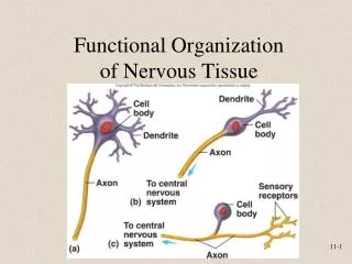
Neurons (Nerve Cells) • Structural units of the nervous system • Receive stimuli and transmit action potentials • Long-life • Amitotic • Have a high metabolic rate • Each neuron consists of: • Body • Axon • dendrites
Structure of Neuron • Cell Body (soma or perikaryon) • Contains the nucleus and a nucleolus & usual organelles • Has no centrioles (amitotic nature) • Has well-developed Nissl bodies (rough ER) • Nissl bodies -primary site of protein synthesis • Contains an axon hillock – cone-shaped area from which axons arise
Dendrites • Short, branched cytoplasmic extensions • They are the receptive, or input, regions of the neuron • Electrical signals are conveyed toward the cell body
Axons • Slender processes of uniform diameter arising from the axon hillock • Initial segment: beginning of axon • Axoplasm : Cytoplasm of axon • Axolemma : Plasma membrane of axon • Long axons are called nerve fibers • Usually there is only one unbranched axon per neuron
Axons • Rare branches, if present, are called axon collaterals • Presynaptic (Axon) terminal – branched terminus of an axon • Trigger zone: site where action potentials are generated; axon hillock and part of axon nearest to cell body • AP are conducted along the axons to axonal terminals and release neurotransmitters • AP conduction away from cell body
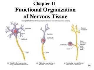
Neuron Classification • Neurons can be classified by structure: • Multipolar • Most common in both CNS & PNS • Single axon, many dendrites (motor neurons and interneurons of CNS) • Bipolar • two processes (one axon and one dendrite) • Are sensory neurons found in the retina, olfactory nerve • Unipolar • single short process extending from cell body • Divides into two branches and functions as both dendrite and axon (sensory neurons , dorsal root ganglia)
Neuron Classification • Neurons can be classified by function: • Sensory (afferent) —transmit impulses from receptors toward the CNS • Motor (efferent) —carry impulses from CNS to muscles and glands • Interneurons (association neurons) Link sensory and motor neurons within CNS • Make up 99% of neurons in body
NERVOUS SYSTEM CELL TYPES • NEUROGLIA (Glial cells) • Supporting cells • Surround neurons • Non-conducting • 6 types • 2 in PNS • 4 in CNS Astrocyte Oligodendrocytes Microglia Ependymal Cells Satellite Cells Schwann Cells
1) Astrocytes In CNS only Anchor neurons to capillaries Regulate what substances reach the CNS from the blood (blood-brain barrier) Regulate extracellular brain fluid composition Pick up excess K+ Recapture released (recycle) neurotransmitters SUPPORTING CELLS OF THE CNS
2) Ependymal Cells CNS only Line the cavities of the brain and spinal cord Ciliated Circulate the cerebrospinal fluid (CSF) SUPPORTING CELLS OF THE CNS
3) Microglia CNS only Migrate toward injured neurons Specialized macrophages Phagocytize necrotic tissue, microorganisms, and foreign substances that invade the CNS SUPPORTING CELLS OF THE CNS
4) Oligodendrocytes CNS only Wrap extensions around neuron fibers (axons) Form myelin sheath SUPPORTING CELLS OF THE CNS
1) Schwann Cells or Neurolemmocytes PNS only Wrap around axons of neurons in the PNS Forms myelin sheath SUPPORTING CELLS OF THE PNS
2) Satellite Cells PNS only Surround neuron cell bodies Provide support and nutrients to neuronal cell bodies Protect neurons from heavy metal poisons (lead, mercury) by absorbing them SUPPORTING CELLS OF THE PNS
Myelinated and Unmyelinated Axons • Myelinated Axons: Whitish, fatty (protein-lipid), segmented sheath around most long axons • Functions: • Protect the axon • Electrically insulate fibers from one another • Increase the speed of nerve impulse transmission • Formed by Schwann cells in the PNS • In CNS formed by oligodendrocytes • Nodes of Ranvier : Gaps in the myelin sheath between adjacent Schwann cells • Unmyelinated Axons : Schwann cell surrounds nerve fibers but coiling does not take place
Organization of Nervous Tissue • White matter – dense collections of myelinated fibers • Gray matter – mostly nerve cell bodies and unmyelinated fibers • In brain: gray is outer cortex as well as inner nuclei; white is deeper • In spinal cord: white is outer, gray is deeper
Synapse Junction between one neuron and another Where two cells communicate with each other Presynaptic neuron – conducts impulses toward the synapse Postsynaptic neuron – Cell that receive the impulse Most are axo-dendriticor axo-somatic NEURON FUNCTION: The Synapse
Types of Synapses • Electrical Synapses: • Are gap junctions that allow ion flow between adjacent cells by protein channels called Connexons • Not common in CNS • Found in cardiac muscle and many types of smooth muscle Action potential of one cell causes action potential in next cell
Chemical Synapses Most are this type Neurotransmitter released from synaptic vesicles of presynaptic neuron Neurotransmitter binds to receptors on postsynaptic membrane Binding of neurotransmitter to receptorpermeability change in postsynaptic membrane Chemical Synapses
NEUROTRANSMITTERS • Released at chemical synapses • In response to AP Voltage-regulated calcium channels open • Ca2+ diffuse into presynaptic terminal • And causes synaptic vesicles to fuse with presynaptic membrane • This fusion releases neurotransmitter into the synaptic cleft via exocytosis
When bound to receptors on postsynaptic neuron, the neurotransmitter can either excite or inhibitthe postsynaptic neuron NEUROTRANSMITTERS
Resting neurons maintain a difference in electrical charge inside and outside cell membrane = RESTING MEMBRANE POTENTIAL (RMP) The inside of the resting neuron is negatively charged, the outside is positively charged. Concentration of K+ higher inside than outside cell Na+ higher outside than inside RMPs vary from -40 to -90mV in different neuron types Resting Membrane Potential
When bound to receptors on the postsynpatic neuron membrane: Causes the opening of positive ion channels Sodium ions enter rapidly RMP becomes more positive This positive change in the RMP is called depolarization This brings the neuron closer to firing EXCITATORY NEUROTRANSMITTERS
DEPOLARIZATION • A positive change in the RMP • Caused by influx of positive ions • Causes the inside of the cell membrane to become less negative • Depolarization spreads to adjacent areas
When bound to receptors on the postsynaptic membrane: Make the membrane more permeable to negative ions (usually Cl-) As negative ions rush into the neuron, the RMP becomes more negative The negative change in the RMP = hyperpolarization Brings the neuron farther from firing INHIBITORY NEUROTRANSMITTERS
Hyperpolarization • A negative change in RMP • Usually caused by influx of chloride ions • Decreases the likelihood of the neuron firing
Graded or Local Potential • Short changes in the RMP in small regions of the membrane • Can be positive or negative (depolarize or hyperpolarize the membrane) • Alone, not strong enough to initiate an impulse • summate or add onto each other • Together, can trigger a nerve impulse (action potential)
EPSP (Excitatory Postsynaptic Potential) When depolarization occurs, response is stimulatory & graded potential is called EPSP Binding of a neurotransmitter on the postsynaptic membrane more positive RMP, reaches threshold (depolarization occurs) producing an action potential and cell response POSTSYNAPTIC POTENTIALS
IPSP (Inhibitory Postsynaptic Potential) When hyperpolarization occurs, response is inhibitory & graded potential is called IPSP Binding of the neurotransmitter on the postsynaptic membrane more negative RMP (hyperpolarization) Decrease action potentials by moving membrane potential farther from threshold POSTSYNAPTIC POTENTIALS
40 to 50 Known Neurotransmitters Acetylcholine (ACh) Norepinephrine (NE) GABA Dopamine Serotonin Types of Neurotransmitters
Action Potential = Nerve Impulse Consists of: Depolarization Propagation Repolarization Action Potential
Action Potential • If depolarization reaches threshold (usually a positive change of 15 to 20 mV or more), an action potential is triggered • The positive RMP change causes electrical gates in the axon hillock to open • Sudden large influx of sodium ions causes a reversal in the membrane potential (becomes approx. 100mV more positive) • Begins at the axon hillock and travels down the axon
Types of Ion Channels • Chemically gated channels – open with binding of a specific neurotransmitter • Voltage-gated channels – open and close in response to membrane potential Chemically Gated (on dendrite or soma) Voltage Gated (on axon hillock and axon)
Movement of the action potential down the axolemma voltage-gated sodium channels open in response to positive RMP change Propagation
Restoration of the RMP back to it’s negative state A repolarization wave follows the depolarization wave 3 factors contribute to restoring the negative RMP: Sodium (Na+) gates close (it no longer enters) Potassium (K+) gates open, potassium rushes out Sodium/potassium pump kicks in REPOLARIZATION
An active process: requires cellular energy Actively pumps 3 sodium (Na+) ions out of the cell and 2 potassium (K+) ions in Potassium leaks back out THE SODIUM/POTASSIUM PUMP
Period of time when electrical sodium gates are open From beginning of action potential until near end of repolarization No matter how large the stimulus, a second action potential cannot be produced ABSOLUTE REFRACTORY PERIOD
Relative Refractory Period • The interval following the absolute refractory period when: • Sodium gates are closed • Potassium gates are open • Repolarization is occurring • A stronger-than-threshold stimulus can initiate another action potential
SUMMATION BY POSTSYNAPTIC NEURON • A single EPSP cannot induce an action potential • EPSP’s can add together or SUMMATE to initiate an action potential • Spatial Summation • Large numbers of axon terminals stimulate the postsynaptic neurons simultaneously
Temporal Summation One or more presynaptic neurons transmit impulses in rapid fire succession SUMMATION BY POSTSYNAPTIC NEURON
An action potential is an “all or none” phenomenon When threshold is reached, the action potential will occur completely If threshold is not reached, the action potential will not occur at all ALL-OR-NONE RESPONSE
Occurs only in myelinated axons Depolarization wave jumps from one node of Ranvier to the next Results in faster nerve impulse transmission SALTATORY CONDUCTION
A nerve impulse in the presynaptic neuron causes release of neurotransmitter into synaptic cleft Neurotransmitter binding to receptors on postsynaptic neuron dendrite or soma cause certain chemically gated ion channels to open If Na+ channels open: Rapid influx of Na+ ions (depolarization) A small positive graded potential occurs (EPSP) If RMP changes in a positive direction by 20mV (or reaches the threshold), voltage gated sodium channels in the axon hillock open Sodium rushes in at the axon hillock resulting in an action potential As the positive ions get pushed down the axon, more voltage gated sodium channels open and the depolarization continues down the axon (propagation) The process of restoring the negative RMP begins immediately following the depolarization wave (repolarization) SUMMARY OF EVENTS