Download
1 / 47
570 likes | 764 Vues
Management of common joint dislocation for medical practitioner.

E N D
DISLOCATION Prepared by: DR ahmadzhafir bin zulkifli Mentor: DR mohdfirdaus bin azizi Supervisor: MR ahmadmahyuddin bin Mohamed Shoulder dislocation Hip dislocation Elbow dislocation Knee dislocation
SHOULDER ANATOMY • Shoulder region: glenohumeral joint, acromioclavicular joint, coracoclavicular joint • Ball and socket synovial joint • Bones • Rotator cuff muscles • Ligaments • Capsule • Stability: anterior and posterior
ANTERIOR SHOULDER DISLOCATION • 90% of shoulder dislocation, why? • Mech: abduction, external rotation, extension • c/o: shoulder pain, deformity, swelling, loss of function • Important: axillary nerve • XRAY • Management: CMR, immobilization
Scapula Y view Recurrent shoulder dislocation: Hill-Sachs lesion: posterolateral humeral head compression fracture 2. Bankart lesion: detachment of the anterior inferior labrum
Hippocratic method • Gently increasing traction is applied to the arm with the shoulder in slight abduction • Assistant applies firm counter traction to the body (a towel slung around the patient’s chest, under the axilla) Source: Reichman EF: Emergency Medicine Procedures Second Edition, McGraw-Hill
CMR: Traction – Metsen’s traction counter traction technique • Patient in supine • Place sheet around the chest and also around the assistant’s waist for countertraction • Flex elbow 90 degree then provide traction at the arm Source: Comparison of four different reduction methods for anterior dislocation of the shoulder, Journal of Orthopaedic Surgery and Research May 2015
CMR: Stimson’s technique • Patient is left prone with the arm hanging over the side of the bed • After15 or 20 minutes the shoulder may reduce Source: AOTrauma Reduction of the glenohumeral joint
Milch technique • Patient lie supine • Pulls the arm first to a 90°abduction • Then slow external rotation to 90° • Nearly painless Source: Medscape Shoulder Dislocation Reduction Technique
Spaso method • Patient in supine • Hold arm around the wrist or the distal forearm • Gentle vertical traction • While on traction, external rotate the shoulder Source: Comparison of four different reduction methods for anterior dislocation of the shoulder, Journal of Orthopaedic Surgery and Research May 2015
CMR: Kocher’s method • Elbow is flex 90° and held close to the body • No traction • Arm is slowly rotated 75 degrees externally • Point of the elbow is lifted forward • Arm is rotated internally • **Not recommended: fracture proximal humerus and neurovascular risk
Post reduction mx • Complication!! check neurovascular: axillary, median, ulnar, radial, distal pulses • Arm sling 3 weeks (less than 30 yo) to rest shoulder, physio elbow and fingers, 1 week for those who prone to have stiffness • Recurrent dislocation, athletes -> surgery
Complication • Early: • Rotator cuff tear: unable to abduct • Axillary nerve injury • Vascular injury • Fracture-dislocation • Late: • Stiffness • Unreduced dislocation • Recurrent dislocation
POSTERIOR SOULDER DISLOCATION • Less common, why? • Suspected in: seizure, electroconvulsive therapy, direct blow from front • Mech: Internally rotated, adducted • Clinical: internally rotated arm vs XRAY AP view • Dx frequently missed – essential lateral view • Mx post reduction: same as anterior
CMR technique • Pulling on the arm with the shoulder in adduction • Wait few minutes for head of the humerus to disengage • Gentle external rotate • Further mx: • If stable can put in armsling • If unstable: held in airplane splint for 3-6 weeks to allow for posterior capsule healing • Physiotherapy ROM shoulder joint
Apprehension test: positive when the shoulder is passively manipulated into abduction, extension and external rotation, the patient tenses up and anxiously resists further movement
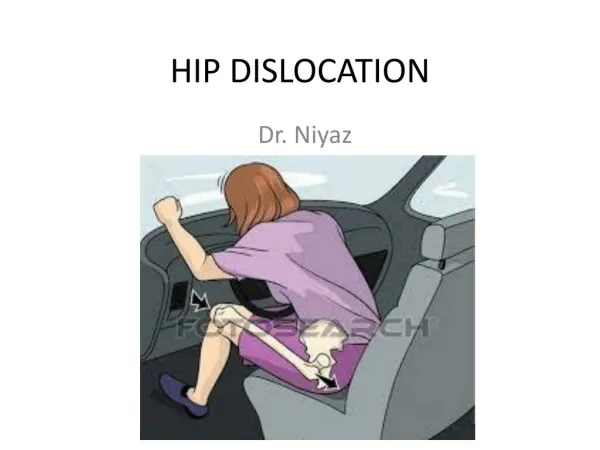
HIP DISLOCATION • Great amount of force: thus fracture is common • 3 types: anterior, posterior, central • Posterior dislocation more common, WHY? • ANY TYPE IS EMERGENCY! • Posterior dislocation: flexed, adducted, internally rotated • Check sciatic nerve? • Complication: AVN, acetabular wall fracture, femoral head fracture, sciatic nerve injury, osteoarthritis ) – CT hip post reduction
CMR technique • Assistant steadies the pelvis • Surgeonapply traction in the line of the femur as it lies (usually in adduction and internal rotation • Then gradually flexes the patient’s hip and knee to 90 degrees, maintaining traction throughout. • At 90 degrees of hip flexion, traction is steadily increased and sometimes a little rotation (either internal or external) is required to accomplish reduction. • Another assistant can help by applying direct medial and anterior pressure to the femoral head through the buttock. • A satisfying ‘clunk’ terminates the manoeuvre. • Telescoping test
Post CMR mx • Immobilize: apply traction and maintain it for a few days • Movement and exercises are begun as soon as pain allows • Continuous passive movement (CPM) machines useful • Excessive hip movements are avoided to allow healing of the capsule and ligaments • After 2 weeks, allow crutches ambulation, non weight bearing at affected side • “Hip protection” 6-12 weeks
ELBOW DISLOCATION • Second most common dislocation • Paediatrics age group • Posterior or Posterolateral (90%) – often with fracture of the body processes • Mech: FOOSH + elbow extension • Clinical: patient supports his forearm with the elbow in slight flexion, deformity • Vascular and nerve damage • Xray: may have fractures
Elbow dislocation, mx: • Uncomplicated: reduction under anaesthasia, check ROM+ neurovascular, collar and cuff 90 degree flexion • Unstable: • Corocoid process fracture • Medial epicondyle fracture • Head of radius fracture • Olecranon fracture • Side-swipe injury • Persistent instability
Elbow dislocation, complication: • Early: brachial artery, ulnar nerve • Late: • Stiffness • Myositis ossificans • Unreduced dislocation • Recurrent dislocation • Osteoarthritis
KNEE DISLOCATION • The knee can be dislocated only by considerable violence, as in a road accident. The cruciate ligaments and one or both lateral ligaments are torn. • Clinical: severe bruising and swelling • popliteal artery?, Common peroneal nerve? • Compartment syndrome • Xray: may reveal fracture • Mx: • 1. urgent reduction under anaesthasia (pulling), careful to avoid hyperextension, why? • 2. splinting
Complication: • Early: vascular injury, nerve damage • Late: joint stiffness, joint instability
REFERENCES • Apleyand Solomon’s Concise System of Orthopaedics and Trauma 4th Edition • Apley’s System of Orthopaedics and Fractures 9th Edition • Radiopaedia.org • OrthoInfo by American Academy of Orthopaedic Surgeon • ORTHOBULLETS • Medscape • Journal of Orthopaedic Surgery and Research