Download
1 / 36
360 likes | 523 Vues
The Spinal Cord. Chapter 13. I. Gross Anatomy of the Spinal Cord. ~18 inches long by ~1/2 inches wide See handout for diagram of anatomy Find and know these terms: Posterior medial sulcus Anterior median fissure Cervical & lumbar enlargements Conus medullaris

E N D
The Spinal Cord Chapter 13
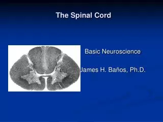
I. Gross Anatomy of the Spinal Cord • ~18 inches long by ~1/2 inches wide • See handout for diagram of anatomy Find and know these terms: • Posterior medial sulcus • Anterior median fissure • Cervical & lumbar enlargements • Conusmedullaris • Filumterminale (“terminal thread”) • Dorsal root ganglion • Dorsal & ventral roots • Spinal nerves • Caudaequina (“horse tail”)
Spinal Meninges • a series of specialized membranes that provide physical stability and shock absorption • Blood vessels lie within the layers to deliver oxygen and nutrients. • are continuous with the cranial meninges surrounding the brain
Bacterial/viral infection can cause meningitis • inflammation of the meninges • can disrupt circulation of cerebrospinal fluid (CSF) • can be deadly • Meningitis & college… should you get vaccinated?
Three meningeal layers • Dura Mater (“hard mother”) • tough, fibrous, outermost covering • padded from vertebrae by adipose tissue in the epidural space • Injecting an anesthetic into this space will affect spinal nerves near the injection site. • May be used in the lower lumbar or sacral region during childbirth.
Arachnoid (“spider”) • middle layer • encloses the subarachnoid space • contains a network of collagen and elastic fibers (arachnoidtrabeculae) • filled with CSF - shock absorption - diffusion of nutrients and wastes • Spinal taps occur here - withdrawal of CSF with a hollow needle in the lower lumbar region - used to diagnose problems like meningitis
Pia Mater (“delicate mother”) • innermost layer • contain blood vessels that service the spinal cord • bound firmly to the underlying neural tissue
II. Sectional Anatomy of the Spinal Cord • See handout for diagram of anatomy • Organization of Gray Matter • Reminder: gray matter= unmyelinated neuron cell bodies • Functional groups: • Sensory nuclei • Motor nuclei
Organization of White Matter • Reminder: white matter = myelinated axons • Each side is divided into three regions called columns or funiculi: • Posterior white columns • Anterior white columns • Lateral white columns • Each column contains tracts (bundles of axons): • Ascending tracts – sensory info • Descending tracts – motor info
III. Spinal Nerves • Each spinal nerve is covered by a series of connective tissue layers • Epineurium- outermost layer • Perineurium • middle layer • surrounds bundles of axons (fascicles) • contains blood vessels • Endoneurium • innermost layer • surrounds individual axons
See handout for diagram of anatomy • Dermatomes: • a specific region of the body surface that is monitored by a pair of spinal nerves • Damage or infection of a spinal nerve will produce a loss of sensation in the specific skin region • example: Shingles (ask Ms. Schroeder!)
Nerve Plexuses (“nerve braid”) • complex, interwoven network of nerves • Three major plexuses: • Cervical plexus- spinal nerves C1-C5 • Brachial plexus- spinal nerves C5-T1 • Lumbosacral plexus- T12-S4
The Brain Chapter 14
I. Introduction to the Organization of the Brain • Contains ~98% of body’s neural tissue • Weighs ~3lbs & has a volume ~1200 cm3 • Can vary from 750 cm3 – 2100 cm3 • Men’s brains are ~10% larger than females • No correlation exists between size & intelligence
Preview of Major Regions & Landmarks • Cerebrum • Largest in size of all the regions • Consists of paired cerebral hemispheres • Conscious thoughts, sensations, intellect, memory & complex movements originate here • Surfaces are highly folded and covered by neural cortex, a superficial layer of gray matter (cerebral cortex)
Cerebellum • Second largest in size • Also divided into hemispheres, partially hidden by the cerebral hemispheres • Adjusts ongoing movements on the basis of comparisons between new and old sensations
Diencephalon • Links the cerebral hemispheres and the brain stem • Left & right thalamus • Contain relay and processing centers for sensory information • Hypothalamus • Floor of diencephalon • Connected to the pituitary gland • Involved with emotions, autonomic function & hormone production
Brain stem • Contains a variety of important processing centers that relay information headed to or from the cerebrum or cerebellum • Includes: • Mesencephalon • Contain sensory nuclei that process visual & auditory information & control reflexes for these stimuli
Pons (“bridge”) • Connects cerebellum to brain stem • Contains tracts, relay centers & nuclei involved with somatic & visceral motor control • Medulla Oblongata • Connects brain to spinal cord • Relays sensory information to the thalamus and the brain stem • Contains major centers concerned with regulation of autonomic function (heart rate, blood pressure, digestion)
Ventricles of the Brain • Chambers filled with CSF and lined by ependymal cells • CSF continually circulates between the ventricles, central canal & subarachnoid space • 4 chambers: • 1st Lateral Ventricle • 2nd Lateral Ventricle • 3rd Ventricle - in the diencephalon • 4th Ventricle
II. Protection & Support of Brain • Brain is protected by: • Cranial bones • Cranial meninges • same 3 from spinal cord are continuous in the brain w/ some differences • Dura mater has 2 layers w/ the superficial layer fused w/ the periosteum of the cranial bones • No epidural space • Dural layers are separated by a small gap & in some places large channels called dural sinuses
Functions of the Cranial Meninges • Tough, fibrous dural folds hold brain in position • CSF acts as a shock absorber
Cerebrospinal fluid • Produced by ependymal cells in the choroid plexus • ~150 ml of CSF in the nervous system • Entire volume of CSF is replaced every 8 hrs • Blood-brain-barrier