Download
1 / 29
290 likes | 703 Vues
The Spinal Cord. Chapter 13. Functions. The spinal cord with its 31 pairs of spinal nerves serves two important functions. It is the connecting link between the brain and most of the body. It is involved in spinal reflex actions, both somatic and visceral. Basic Anatomy of the Spinal Cord.

E N D
The Spinal Cord Chapter 13
Functions • The spinal cord with its 31 pairs of spinal nerves serves two important functions. • It is the connecting link between the brain and most of the body. • It is involved in spinal reflex actions, both somatic and visceral.
Basic Anatomy of the Spinal Cord • The spinal cord extends caudally from the brain for about 45 cm and has a width of ~14 mm. Its upper end is continuous with the brain (medulla oblongata). The cord is slightly thicker than a pencil. • There are 31 pairs of spinal nerves:8 cervical, 12 thoracic, 5 lumbar, 5 sacral, and coccygeal. The roots of the lumbar and sacral are called cauda equina. • Surrounding and protecting the spinal cord is the vertebral column. • The spinal cord is slightly flattened dorsally and ventrally, with two enlargements-cervical and lumbosacral from which the spinal nerves emerge that innervate the upper and lower limbs.
Basic Anatomy of the Spinal Cord • The cervical enlargement supplies nerves to the pectoral girdle and upper limbs. • The lumbar enlargement supplies nerves to the pelvis and lower limbs. • Inferior to the lumbar enlargement, the spinal cord becomes tapered and conical-conus medullaris. • Filum terminale-slender strand of fibrous tissue that extends from conus medullaris.
Spinal Nerves • There are 8 cervical nerves(C), 12 thoracic(T), 5 lumbar (L), 5 sacral (S), and 1 coccygeal (Co). • Each pair of spinal nerves passes through a pair of intervertebral foramina located between two successive vertebrae. Each spinal nerve caudal to the first thoracic vertebra takes its name from the vertebra immediately preceding it. • The nerves are then distributed to a specific pair of segments of the body. • The spinal cord and the roots of its nerves are protected by the vertebral column, its ligaments, spinal meninges and cerebrospinal fluid.
Spinal Meninges • The outer layer is called dura mater. This is a tough, fibrous memebrane that merges with the filum terminale. • The middle layer, the arachnoid, runs caudally to the S2 vertebral level. This is delicate and transparent. • The innermost is called, pia mater. It is highly vascular and tightly attached to the spinal cord and its roots. • Meningitis-bacterial or viral infection.
Spinal Meninges • Between the dura mater and periosteum of the vertebrae is the epidural space that contains many blood vessels and fat. • Anesthetics can be injected here below the L3 vertebral level, from which it ascends to act upon sensory neurons to help dull pain. This procedure is called caudal block.(epidural block) • Space between dura mater and archnoid-subdural space-no CSF. • Space between arachnoid and pia mater-subarchnoid space-CSF, blood vessels, spinal roots.
Cerebrospinal Fluid • This is a clear watery ultra filtrate solution primarily derived from blood. • The basic mechanism involves an active transport system and passive diffusion into the four ventricles. • The CSF provides a cushion that protects the delicate tissues of the spinal cord. • It is also involved in the exchange of nutrients between the blood and neurons of the brain and spinal cord.
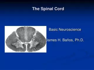
Internal Structure • If the spinal cord is cut in X.S., a tiny central canal is observed, which contains CSF. • There is a dark portion of H-shaped or butterfly shaped “gray matter”, surrounded by a larger area of “white matter”. • The spinal cord is divided into more or less symmetrical halves by a deep groove called the anterior(ventral) median fissure and a median septum called posterior (dorsal) median sulcus. • Extending from the spinal cord are the ventral and dorsal roots of the spinal nerves.
Gray Matter • The gray matter of the spinal cord consists of nerve cell bodies, dendrites and axon terminals(unmyelinated) and neuroglia. It is pinkish-gray color because of a rich network of blood vessels. • The gray matter forms an H shape and is composed of three columns of neurons-posterior, anterior and lateral horns. The projections of gray matter toward the outer surface of spinal cord are called horns. • The two that run dorsally-posterior horns which function in afferent input. The two that run ventrally-anterior horns which function in efferent somatic output. The two that extend laterally-lateral horns. • The nerve fibers that form the cross of the H are known as gray commisure-functions in cross reflexes.
White Matter • The white matter gets its name because it is mainly composed of myelinated nerve fibers, and myelin has a whitish color. • The white matter is divided into three pairs of columns or funiculi of myelinated fibers-anterior, posterior, lateral and a commisure area. • The bundles of fibers within each funiculus are divided into tracts called fasciculi. • Ascending tracts-sensory fibers carry impulse up the spinal cord to the brain. • Descending tracts-motor neurons transmit impulse from the brain down the spinal cord.
Spinal Nerves • A series of connective tissue layer surrounds each spinal nerve. • Epineurium-outermost layer, consists of a dense network of collagen fibers. • Perineurium-extend inward from th epineurium, dividing the nerve into a series of compartments. • Endoneurium-delicate connective tissue fibers.
Ventral and Dorsal Roots • In the vicinity of the cord, each spinal nerve divides into a ventral (anterior, motor) root and a dorsal (posterior, sensory) root. • Ventral roots contain mostly efferent nerve fibers and convey motor information. • Dorsal roots contain afferent nerve fibers and convey sensory information. • The axons of motor neurons whose cell bodies are located within the CNS in the ant. Horn emerge from the spinal cord to form ventral roots (motor). • Groups of sensory neurons , whose axons make up the dorsal roots lie outside the cord in the dorsal root ganglia or spinal ganglia of the PNS.
Peripheral distribution of Spinal Nerves • A typical spinal nerve has a white ramus(this contains myelinated axons), and a gray ramus (unmyelinated fibers that innervate glands and smooth muscles in the body wall or limbs) • A dorsal ramus(providing sensory and motor innervation to the skin and muscles of the back), and a ventral ramus (supplying the ventrolateral body surface, structures in the body wall and the limbs). • Each pair of nerves monitors a region of the body surface called a dermatome.
Nerve Plexuses • A complex, interwoven network of nerves is a nerve plexus. • The three large plexuses are the cervical plexus, the brachial plexus and the lumbosacral plexus. The latter can be further divided into the lumbar plexus and the sacral plexus.
Functional Roles of Pathways of CNS • Each pathway is composed of organized sequences of neurons. • Upper motor neurons in the brain influence the activity of lower motor neurons in the cranial and spinal nerves. • Some neurons have long axons that terminate in processing centers-called nucleus, ganglion, gray matter of spinal cord or cortex of the brain.
General Somatic Efferent (Motor) System • The brain exerts active influences on the activity of skeletal muscles through descending motor pathways that make up the upper motor neurons. • These originate from the cell bodies in the cerebral cortex and brainstem. • These act by regulating and modulating the activity of the lower motor neurons of the cranial and spinal nerves.
Lower Motor Neurons • These include alpha and gamma motor neurons. • Alpha motor neurons have their cell bodied in their CNS. Their axons course through cranial and spinal nerves and terminate on the motor end plates of skeletal muscle fibers (extrafusal muscle fibers). Involved in stretch reflex. • Gamma neurons also have cell bodied within the CNS. Their axons pass through cranial and spinal nerves to innervate the intrafusal muscle fibers inside the neuromuscular spindles. Involved in the gamma motor neuron reflex.
Lower Motor Neurons • These are the only neurons that innervate the skeletal muscle fibers, they function as the final common pathway, the final link between the CNS and skeletal muscles. • Axons are located both in the cranial and spinal nerves. • Those in cranial nerves innervate the skeletal muscles associated with the movements of the eyes, tongue, chewing, swallowing, vocalizing. • These are influenced by two sources: sensory receptors that are integrated into reflexes and upper motor neurons from the brain that form the “voluntary descending pathways”.
Upper Motor Neurons • This is entirely located in the CNS.
Sensory Pathways • Some of these have sequences that are made of three neurons. • They may be called first, second and third order neurons. • A first-order neuron extends from sensory receptor to CNS. • A second-order neuron extends from the spinal cord or brainstem to nucleus in the thalamus. • A third-order neuron extends from the thalamus to a sensory area of the cerebral cortex.
A critical feature of many pathways is that they cross over or decussate. By knowing where a pathway crosses over, a physician can use this information to help locate the site of an injury in the CNS. • Example is touch-pressure pathway that decussates in the medulla oblongata.
Tracts • Many tracts are named after their nuclei of origin, their termination as well as their location in the spinal cord (eg. Lateral spinothalamic tract). • Anterolateral System: this consists of the lateral spinothalamic tract, spinoreticulothalamic tract and anterior spinothalamic tract. This involves the general sensatins of pain, temperature and light touch. • Posterior-column medial lemniscus pathway.
Reflexes • A reflex is a predictable involuntary response to a stimulus. • A reflex involving the skeletal muscles is called a somatic reflex. • A reflex involving responses of smooth muscle, cardiac muscle, or a gland is a visceral reflex.
Classification of Reflexes • Reflexes are classified according to : • 1) their development : innate and acquired • 2) site of information processing: cranial and spinal reflexes. • 3)nature of resulting motor response : somatic and visceral reflexes. • 4) the complexity of the neural circuit : monosynaptic and polysynaptic reflexes.
Spinal Reflexes • A reflex always starts with a sensory neuron and ends with a motor neuron. • In a monosynaptic (one synapse, two neuron) reflex arc, the sensory and motor neurons synapse directly. • More often, however, one or more interneurons synapse with the sensory and motor neurons in a polysynaptc reflex arc. • Most reflex actions never travel any higher than spinal cord. Some maybe to brain stem.
Types of Reflexes • Stretch (Myotatic Reflex)-monosynaptic reflex arc. Maintains erect posture. Eg. Knee-jerk or patellar reflex. Ipsilateral-response and stimulus on same side. • Gamma Motor Neuron Reflex Arc-this acts to smooth out the movements of muscle contractions or to sustain the contraction of a muscle.
Types of Reflexes • Plantar Reflex: this clinically tests the integrity of the spinal cord from L4 to S2. It is tested by drawing a blunt instrument down the lateral aspect of the sole(plantar surface) of the foot. A normal response is a curling or downward flexion of the foot. • Withdrawal reflex arc: involves sensory receptors, afferent neurons, interneurons, alpha motor neurons, skeletal muscles.
Some Diagnostic Reflexes • Abdominal reflex • Achilles Reflex • Biceps Reflex • Babinski’s reflex • Hoffmann’s reflex • Patellar Reflex • Plantar reflex • Triceps reflex