Download
1 / 33
650 likes | 1.46k Vues
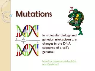
MUTATIONS. Fitness of Mutations. The fitness of a mutation describes its value to the survival and reproductive success of the organism. A mutation may turn out to be: Lethal : Many mutations are lethal and embryos are non-viable .

E N D
Fitness of Mutations • The fitness of a mutation describes its value to the survival and reproductivesuccess of the organism. • A mutation may turn out to be: • Lethal: Many mutations are lethal and embryos are non-viable. • Harmful: Non-lethal mutations, e.g. Down syndrome and sickle cell disease, may be expressed as effects that lower fitness. • Silent (neutral): Most point mutations are probably harmless, with no noticeable effect on the phenotype. • Beneficial (useful): Occasionally mutations may be useful, particularly in a new environment, e.g. insecticide resistance in insects, antibiotic resistance in bacteria.
Location of Mutations Gametic Mutations Somatic Mutations Mutation Mutation Sperm Sperm • The location of a mutation determines whether or not it will be inherited. • Most mutations occur in somaticcells and are not inherited. • Gameticmutations occur in the cells of the gonads (which produce sperm and eggs) and may be inherited. Egg Egg Fertilisation Mutation Mutation Cleavage. Prior to implantation Fetus Baby Cells of tissues affected by the mutation Gametic mutations are inherited and occur in the testes of males and the ovaries of females. Somatic mutations occur in body cells. They are not inherited but may affect the person during their lifetime.
Neutral Mutations Normal DNA • Neutral mutations are hard to detect because they produce little or no change in the phenotype. • They may have little or no effect on the survival of an organism or its ability to reproduce. • They may be the result of a ‘same-sense’ mutation where a change in the third base of a codon still codes for the same amino acid. mRNA Amino acids Amino acid sequence from the non-mutated DNA forms a normal polypeptide chain Mutation: Substitute C instead of T Mutant DNA mRNA Amino acids Despite the change in the last base of a triplet, the amino acid sequence is unchanged
Italy Limone Lake Garda Brescia Verona Beneficial Mutation Example • Tolerance to high cholesterol levelsin humans • In the small village of Limone, about 40 villagers have extraordinarily high levels of blood cholesterol, with no apparent harmful effects on their coronary arteries. • The village has a populationof 980 inhabitants and was,until recently, largely isolatedfrom the rest of the world, withsheer cliffs behind the village,the lake in front of them,and no road access. The village of Limone, on the shore of Lake Garda, Italy
Beneficial Mutation Example • The 40 villagers possess a point mutation which alters the protein produced by just one amino acid. This protein is ten times more effective at mopping up excess cholesterol. • No matter how much excess cholesterol is ingested, it can always be disposed of. • All carriers of the mutation are related and have descended from one couple who arrived in Limone in 1636. • Generally, the people of Limonelive longer and show a highresistance to heart disease. High blood cholesterol and dietary fat are implicated in the formation of plaques in the coronary arteries and in the development of cardiovascular disease.
Point Mutations 2 • As a reference for the following screens, the diagram below illustrates the transcription and translation of DNA without a point mutation. Original Unaltered Code Original DNA Transcription mRNA Translation Amino acids Amino acid sequence forms a normal polypeptide chain
Mutation: Substitute T instead of C Polypeptide chain with wrong amino acid Missense Substitution • A single base is substituted by another. • Usually results in coding for a new amino acid in the polypeptide chain. • If the third base in a triplet had been substituted, the resulting amino acid may not be altered (due to degeneracy in the code). Original DNA Mutant DNA mRNA Amino acids
Mutation: Substitute A instead of C Mutated DNA creates a STOPcodon which prematurely ends synthesis of the polypeptide chain Nonsense Substitution • A single base is substituted by another. • This results in a new triplet that does not code for an amino acid. • The resulting triplet may be an instruction to terminate the synthesis of the polypeptide chain. Original DNA Mutant DNA mRNA Amino acids
First base Sickle Cells containing mutant hemoglobin (less soluble) Normal Red Blood Cells containing normal hemoglobin (soluble) DNA Codes for the 1st amino acid Beta (β) chain Alpha (α) chain Sickle Cell Mutation • The mutation responsible for causing sickle cell disease is a point substitution mutation. Hemoglobin molecules are made up of 2 α-chains and 2 β-chains linked together Hemoglobin clusters together to form fiber, which deform the red blood cells into a sickle shape β-Chain hemoglobin Normal base: T Substituted base: A The sickle cell mutation involves the substitution of one base for another in the HBB gene, causing a single amino acid to be altered.
Sickle Cell Disease Gene location: Chromosome 11 HBB p q • Synonym: Sickle cell anemia • Incidence: Most common in people of African ancestry. • WestAfricans: 1%(10-45% are carriers) • WestIndians: 0.5% • Gene type: Autosomal recessive mutation (HBB) on chromosome 11 which results in the substitution of a single nucleotide in the HBB gene coding for the beta chain of hemoglobin. Normal red blood cells Sickle-cell Photo Defiers.com
Sickle Cell Disease • Symptoms include the following: • Pain, ranging from mild to severe, in the chest, joints, back, or abdomen • Swollen hands and feet • Jaundice • Repeated infections, particularly pneumonia and meningitis • Kidney failure • Gallstones (at an early age) • Strokes (at an early age) • Anemia.
Mutation: Insertion of C Large scale frame shift results in a new amino acid sequence. The resulting protein is unlikely to have any biological activity. Reading Frame Shift by Insertion • A single base is inserted, upsetting the reading sequence for all those after it. • A reading frame shift results in new amino acids in the polypeptide chain from the point of insertion onwards. • The resulting protein will be grossly different from the one originally encoded (it is most likely to be non-functional). Original DNA Mutant DNA mRNA Amino acids
Mutation: Insertion of C Mutation: Deletion of C Altered chain which may or may not produce a protein with biological activity Partial Reading Frame Shift • A single base is inserted and another is deleted at a different location. This causes a localised frameshift. • The amino acid sequence between these points changes. • Depending on how many amino acids are affected, the resulting protein may have some biological activity. Original DNA Mutant DNA mRNA Amino acids
Cystic Fibrosis Mutation • The mutation causing 70% of cystic fibrosis cases is a gene mutation (delta F508) involving a triplet deletion. Base 1630 CFTR protein Part of the DNA sequence in the CFTR gene This triplet codes for the 500th amino acid The 508th triplet is absent in the mutant form Cl- Cl- Cl- Water Cl- Outsidethe cell Outsidethe cell Cellmembrane Cl- Cl- Cellcytoplasm Cellcytoplasm Cl- Cl- Cl- Cl- Cl- Normal CFTR protein regulates chloride transport across the membrane Mutant CFTR protein cannot regulate chloride transport. Chloride ions to remain in the cell and water enters the cell
Cystic Fibrosis • Synonyms: Mucoviscidosis, CF • Incidence: Varies with populations: • Asians: 1 in 10 000 • Caucasians: 1 in 20-28 are carriers • Mutation type: Autosomal recessive. Over 500 different recessive mutations of the CFTR gene have been identified: • deletions, missense, nonsense, terminator codon p q CFTR Gene location Chromosome 7
A classic example Sickle-cell anemia: hemoglobin B gene (HBB) Aaheterozygoes partially protected against malaria But aahomozygotes suffer adverse health effects Heterozygote advantage -- but only where malaria is present See: anthro.palomar.edu/synthetic/synth_4.htm
humans and chimpanzees differ in disease outcomes and susceptibility in all 7 4 / 7 are associated with differences in diet between the two species What are the biggest public health concerns? Heart disease Diabetes Infectious disease Cancer Obesity Stroke Neurodegenerative disease Thus, the chimpanzee genome holds important clues to understanding the genetic basis for these diseases in humans.
Block Mutations: Deletion Chromosome rejoins • Break occurs at two points on the chromosome and the middle piece falls out. • The two ends rejoin to form a chromosome deficient in some genes. • Alternatively, the end of a chromosome may break off and be lost. Break Segmentis lost Break Genes Step 1 Step 2 Step 3
Deletion Example Tip deletion Mid-segment deletion • Human chromosome 1 shows two forms of deletion. • These may involve deletion of either a chromosome tip (left) or a middle segment with the tip rejoined (right). • Deletions involving small amounts of chromosomal material underlie several disorders, including: • Cri-du-chat syndrome • Prader-Willi syndrome • Angelman’s syndrome • Deletions of large amounts of chromosomal material areusually lethal. Before After Before After Tip rejoins 1 1 Lost Lost 1 1
Block Mutations: Translocation Segmentremoved • Translocation involves the movement of a group of genes between different chromosomes. • A piece of one chromosome breaks offand joins on to another chromosome. • The result is a chromosome deficient in genes and one with too many genes. Segmentsjoin Break Genes Step 1 Step 2 Step 3
Translocation Example Before translocation After translocation • Translocation can occur between human chromosomes 9 and 22. • The tips of the two chromosomes are exchanged. • This is the translocation observed in chronic myeloid leukemia. 22 22 The tips of the chromosomes swap 9 9
Block Mutations: Inversion • The middle piece of the chromosome falls out, rotates through 180°, and then rejoins. • There is no loss of genetic material. Break Segmentrejoins Break Segmentrotates 180° Genes Step 1 Step 2 Step 3
Inversion Example Normal Inversion • A segment of human chromosome 2 is inverted (caused by looping of the chromosome) • All inversions cause abnormalities during meiosis and affect the viability of the gametes produced. • However, if a combination of genes within an inversion is desirable, they can act as a supergene and can confer a selective advantage. Flip 2 2
Block Mutations: Duplication • A segment is lost from one chromosome and is added to its homologue. • The chromosome on the left was the 'donor' of the duplicated piece of chromosome. • The chromosome with the duplication will become incorporated into a gamete, which may later contribute to an embryo. Joins on to homologous chromosome Segmentremoved Break Genes Step 1 Step 2 Step 3
Duplication Example Normal Duplication • A segment of human chromosome 9 is duplicated. • A segment is taken from its homologue and inserted to produce double copies of some genes. • Some genes may be disrupted. • Gene duplications can have evolutionary significance. Example: The alpha and beta chains of hemoglobin arose following a duplication event 500 million years ago. Duplicatesegment Identicalsegment A segment is tansferred from one chromosome into its homologue 9 9
Maternal Age Effect • Many aneuploidies show a ‘maternal age effect’, where incidence increases with the age of the mother. 1 in 46 Maternal age (years) Incidence per 1000 live births < 30 30 - 34 35 - 39 40 - 44 > 44 < 1 1 - 2 2 - 5 5 - 10 10 - 20 1 in 100 Example:Down syndrome is 100 times more likely in children of mothers over 45 years, than in those of mothers less than 19 years. Estimated rate of Down Syndrome (per 1000 births) 1 in 880 1 in 2 300 1 in 290 Maternal age in years
Causes of Maternal Age Effect • Maternal age effect probably arises because: • All eggs are present at birth but are suspended in their development in early prophase until puberty. • A woman, on average, will produce about 400 eggs in her lifetime (12 per year). • Therefore, by the end of her reproductive life, the egg cells that remain are old and there is a greater chance that errors in meiosis will occur. • A similar, though less marked effect is exerted by the age of the father. Sperm from older men have a slight tendency to be deficient in chromosomes Older egg cells are more prone to faulty meiosis
Autosomal trisomics Trisomy 13 .......... 42 Trisomy 18 .........100 Trisomy 21 ......1041 Other abnormalities Total .......2133 Conceptions 1 000 000 Live births 850 000 Chromosome abnormalities 75 000 With chromosome abnormalities 5165 Perinatal deaths 17 000 Spontaneous miscarriages 150 000 Other causes 75 000 Children 833 000 Trisomics .......39 000 XO .................13 500 Triploids .........12 750 Tetraploids .......4500 Others ..............5250 Sex chromosome aneuploidies Male ..............1 427 Female .............422 The Fate of Conceptions • For every million conceptions that occur, a significant number have genetic abnormalities and fail to develop into a completely normal child: